Sadia Batool, Saba Saba*, Atia Iqbal, Azka Naveed and Afshan Zia
Department of Microbiology and Molecular Genetics, The Women University Multan, Pakistan
* Corresponding Author:[email protected]
Multi-drug resistant (MDR) bacterial infections pose a major threat to global health. The emergence of antibiotic resistance is due to the overuse and misuse of antibiotics. To overcome this problem, phytochemicals extracted from medicinal plants present an attractive alternative. This study was designed to evaluate the antibacterial and antibiofilm activities of Moringa oleifera leaves extracts against human pathogens. Moringa oleifera leaves were collected and their extracts were prepared in methanol, ethanol, water, and dimethyl sulfoxide solvents. Human pathogenic bacteria were isolated from the urine, sputum, and blood samples of patients from a tertiary care hospital in Lahore. Bacterial isolates were characterized based on their morphological, biochemical, and physiological characteristics. Antibacterial activity of antibiotics was checked through the disc diffusion method. Furthermore, the ability of bacterial strains to form biofilms was observed using qualitative ring test and quantitative microtiter plate assay. Bacterial strains were identified as Escherichia coli, Pseudomonas aeruginosa, and Klebsiella pneumoniae. Antibacterial activity of Moringa oleifera leaves extracts, checked by agar well diffusion assay, displayed maximum inhibitory effect (25 mm) in aqueous extract against the strain E2. All of the bacterial strains were found resistant to almost all tested antibiotics, except fosfomycin and amikacin. All bacterial isolates exhibited the potential of biofilm formation. Among all isolates, E2 and E3 bacterial strains appeared as strong slime producers. It was concluded that the significant antibacterial and antibiofilm activity of Moringa oleifera leaves extracts present it as a potential source for novel therapeutic compounds. So, it should be purified and characterized further by using advanced techniques.
Keywords: antibacterial, antibiotic resistance, bacterial strains, biofilm, plant extracts, phytochemicals
Multiple drug resistance (MDR) is a significant challenge in treating any infection. Owing to antimicrobial resistance, there has been a dramatic increase in the mortality rate all over the world [1]. In a survey conducted in 2019, 4.95 million deaths were attributed to antimicrobial resistance and 1.27 million deaths were directly caused by antimicrobial resistant infections [2]. In the past, only a few novel antimicrobials were introduced, while the majority of new drugs introduced in the market were the modifications of already existing drugs [3]. In 2013, it was affirmed that human race is in the ‘post-antibiotic’ era, in which common infections and minor injuries can kill a patient, according to the Centers for Disease Control (CDC), USA [4]. To develop resistance against antimicrobial drugs, bacteria form biofilms in which microbial communities build a complex aggregate of cells and extracellular polysaccharide (EPS) matrix, which make them stand firm against any antimicrobial medicine [5]. A biofilm is a 3D structure that acts as a microbial battlefront. This structure begins to form when a group of microbes’ sense a given surface and adhere to it. Subsequent colonization and production of EPS solidify the structure [6]. Bacteria in a biofilm combie due to biotic (in human tooth, lungs, and intestine of cows) and abiotic factors to form complex masses. Abiotic factors include medical devices, urinary catheters, medical implants, dental devices, contact lenses, and intrauterine and contraceptive devices [7].
Biofilm production and antimicrobial resistance are interconnected. For example, a research conducted on Staphylococcus aureus in 2018 concluded that the biofilm producing S. aureus was concomitant with a higher incidence of antimicrobial resistance, as compared to the microbes that do not produce biofilm [8]. Another study conducted in a tertiary care hospital of Indonesia claimed K. pneumoniae to produce extended spectrum β-Lactamase (ESBLs) and carbapenemase enzymes, which cause to develop higher resistance among K. pneumoniae strains [9].
Plants have been used for the treatment of many ailments in Asia for centuries. According to the World Health Organization (WHO), 80% of the drugs are obtained from natural sources. This is because plant extracts have pharmaceutically important compounds which are considered beneficial in traditional therapies of ailments [10]. Moringa oleifera belongs to the family ‘Moringaceae’ and is also known by some of its traditional names viz drumstick tree, horseradish tree, ben-oil tree, or kelor tree in different regions. M. oleifera originates from the sub-Himalayan tracts of India, Pakistan, Bangladesh, and Afghanistan and is cherished as an edible plant in these countries. For many years, it has been grown in subtropical and tropical climates [11]. It is highly nutritious and provides 7 times more vitamin C, 10 times more vitamin A, 17 times more calcium, 9 times more protein, 15 times more potassium, and 25 times more iron than oranges, carrots, milk, yogurt, bananas, and spinach, respectively [12]. Further, it has anti-cancerous, anti-fungal, antibacterial, antioxidant, cytotoxic, anti-inflammatory, and anti-viral properties [13, 14]. Moreover, ascorbic acid, flavonoids, phenolic acid, and carotenoids are present in M. oleifera leaves. These compounds are pharmaceutically important for anti-oxidation [15]. Through seeding and cutting, M. oleifera is proliferated among all other species to perform diverse functions [16]. The plant has been used as a conventional medicine for infections, skin care, lung problems, stomach problems, anemia, and anxiety in different cultures of the world for many centuries [17]. This study aims to check the inhibitory effect of different solvent extracts of M. oleifera by using agar well diffusion method against pathogenic bacteria.
2.1. Isolation and Characterization of Pathogenic Bacterial Strains
Urine, sputum, and blood samples collected from patients visiting Nishtar Hospital Multan, Pakistan were processed in the laboratory to isolate bacterial strains. Pathogenic isolates were then purified through the quadrant streaking method and characterized morphologically based on colony characteristics including color, appearance, edges, elevations, and transparency. Bacterial strains were characterized biochemically via Gram staining and different biochemical tests including catalase test, oxidase test, IMViC test, and TSI test. For the identification and classification of bacterial strains, Bergey’s manual of systematic bacteriology was followed [18].
2.2. Physiological Characterization of Bacterial Strains
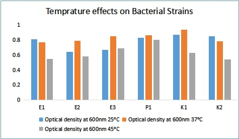
To evaluate the optimum temperature for bacterial growth, 24 h fresh culture was set at 0.5 McFarland standards using the OD600. Further, 100 µl standard culture of bacteria was inoculated in broth media and incubated at different growth temperatures (25°C, 37°C, and 45°C) for 24 hours. OD at 600 nm was measured by using spectrophotometer to evaluate the optimum temperature showing maximum growth.
pH measurements are usually performed using a pH scale calibrated at 25°C (or another specific temperature value), giving acidity measurement or the basics of an aqueous solution. Bacteria require a specific pH to grow perfectly. If the pH changes, bacterial growth is negatively affected. L broth was prepared with pH values 4, 7, and 10 in order to check fortunate pH for the optimal growth of the bacterial sample. The medium was distributed into broth containing tubes. Tubes were inoculated from each pH and 100 µl of standardized bacteria was enriched in these test tubes containing the broth of different pH standards. Each group contained a control tube with only L broth without inoculum (negative control). Incubation was done for a day at 37°C. OD was calculated at 600 nm to evaluate the optimal pH showing maximum growth.
2.3. Disc Diffusion Method
According to the reference to McFarland, 0.5 isolates of bacteria were diluted in normal saline at a concentration of 108 CFU per ml. Therefore, 100µl of suspension was inoculated by swabbing on Mueller Hinton agar plates. Then, the discs of antibiotics were aseptically placed on these inoculated plates, followed by incubation at 37˚C for 24 hours. The zone of inhibition of antibiotics discs was measured in millimeter scale [19].
2.4. Preparation of M. oleifera Extracts
2.5. Antibacterial Activity of Extracts
Agar well diffusion method was used to trace the antibacterial activities of the prepared extracts of leaves. For this purpose, 0.1 ml standardized inoculum of test organisms was uniformly swabbed on the surface of freshly prepared Mueller Hinton agar plates. Wells were punched by cork-borer into each agar plate. Further, 0.2 ml of different extracts of ethanol, methanol, DMSO, and aqueous extract concentrations were placed in every well. Plates were kept in the incubator for incubation at 37˚C for 24 h. The inhibitory effects of extracts against pathogens were reported in millimeter (mm) [20].
2.6. Minimum Inhibitory Concentration (MIC) of Moringa oleifera Extracts
MIC extraction values were determined by following the method with some modifications, while the assay was conducted in 96 well microtiter plates [21]. The first column of the microtiter plate was filled with testing organisms and the remaining wells were filled with LB broth. Double serial dilution was prepared. The extracts solution was diluted by using double serial dilution to obtain final test concentrations of 1000, 500, 250, 125, 62.5, 31.25, 15.63, 7.81, 3.9, and 1.95 ml/ml, respectively. Average values for MIC were measured for test materials.
2.7. Biofilm Formation Assays
Bacterial biofilm formation was checked qualitatively through the following assays, that is, qualitative ring test and slime production test.
2.8. Qualitative Ring Test
All bacterial isolates were monitored for their biofilm forming ability using the ring test. All isolates were cultured on the plate of L agar and kept in incubator at 37°C for 24 h. OD was standardized 0.5 at 600 nm by using spectrophotometer. Small amounts of bacterial culture were dispensed in three pairs of test tubes containing 7 ml of sterile broth. All sets of test tubes were placed in the incubator for distinct time periods (24, 48, and 72 h) at 37˚C. Bacterial solution was carefully aspirated and biofilm rings were stained through 0.1% crystal violet [22].
2.9. Slime Production Test
Slime producing isolates were detected using Congo red agar by following the protocol of Rehman et al. [23]. As a concentrated aqueous solution, the Congo red stain was prepared and sterilized by autoclaving. BHI agar (BRH) was prepared with sucrose and autoclaved for 15 minutes. Congo red stain was added to the sterile sucrose BHI agar at 55°C. Each of the test isolates was isolated on each CRA plate and kept for a day at 98°F. The generation of dark color indicated the production of slime.
3.1. Characterization and Identification of Bacterial Strains
Isolated bacterial strains were characterized morphologically, biochemically, and physiologically.
Isolated bacterial colonies displayed morphological features shown in Table 1.
All of the isolated bacterial strains were Gram negative rods. Biochemically, catalase test, indole test, TSI test, MR test, citrate utilization test, and Hydrogen sulfide test were positive for all strains. Oxidase test was recorded as negative for E1, E2, E3, K1, and K2 strains, while P1 strain was recorded as positive. Voges-Proskauer test was recorded as negative for E1, E2, E3, and P1 strains, while K1 and K2 strains were recorded as positive (Table 2). These results were compared with Bergey’s manual and Escherichia coli, Pseudomonas aeruginosa, and Klebsiella were identified.
Table 1. Morphological Characteristics of Bacterial Strains
|
Bacterial Strains |
Color |
Texture |
Elevation |
Margins |
Opacity |
|
E1 |
white |
Shiny |
Convex |
Irregular |
Translucent |
|
E2 |
Off-white |
Shiny |
Raised |
Smooth |
Opaque |
|
E3 |
Off-white |
Rough |
Flat |
Smooth |
Translucent |
|
P1 |
Green |
Shiny |
Raised |
Irregular |
Translucent |
|
K1 |
Off-white |
Mucoid |
Convex |
Smooth |
Opaque |
|
K2 |
Off-white |
Mucoid |
Raised |
Smooth |
Opaque |
Table 2. Biochemical Characterization of Bacterial Strains
|
Strains |
Bacteria Scientific Name |
Gram stain |
Catalase |
Oxidase |
H2S production |
Indole |
Methyl red |
VP |
Citrate utilization |
TSI |
|
E1 |
Escherichia coli |
Negative rods |
+ve |
-ve |
+ve |
+ve |
+ve |
-ve |
+ve |
+ve |
|
E2 |
Escherichia coli |
Negative rods |
+ve |
-ve |
+ve |
+ve |
+ve |
-ve |
+ve |
+ve |
|
E3 |
Escherichia coli |
Negative rods |
+ve |
-ve |
+ve |
+ve |
+ve |
-ve |
+ve |
+ve |
|
P1 |
Pseudomonas aeruginosa |
Negative rods |
+ve |
+ve |
+ve |
+ve |
+ve |
-ve |
+ve |
+ve |
|
K1 |
Klebsiella pneumoniae |
Negative rods |
+ve |
-ve |
+ve |
+ve |
+ve |
+ve |
+ve |
+ve |
|
K2 |
Klebsiella pneumoniae |
Negative rods |
+ve |
-ve |
+ve |
+ve |
+ve |
+ve |
+ve |
+ve |
|
E1 |
Legends: +ve = positive, -ve = negative
Figure 1. Effect of Temperature Changes on Bacterial Growth
3.2. Physiological Characterization of Bacterial Strains
The growth of bacterial strains was checked at three different temperatures. Overall, bacterial growth was maximum at 37°C in contrast to 25°C and 45°C. Bacterial growth was reduced at high temperature (45°C) in all organisms (Figure 1).
The rise in temperature had an antagonistic relationship with increased growth. The effects of pH changes on the growth of microorganisms was verified. Bacteria were dispensed in medium at distinct values of pH (4, 7, and 9). After 24 h, OD was calculated at 600 nm wavelength. Different conclusions were drawn at distinct pH values (Figure 2). Generally, a pH value of 7 was determined as the best for the better development of living organisms.
3.3. Agar Well Diffusion Assay
Ethanolic, methanolic, DMSO, and aqueous extracts of M. oleifera leaves were added to the wells made on agar plates to evaluate their activities, followed by incubation for 24 h at 37oC. Inhibitory effects against pathogens were calculated in millimeters (Table 3). All pathogenic organisms showed distinct activity against ethanolic, methanolic, DMSO, and water dissolve extracts of M. oleifera (Figure 3).
E2 strain had a greater inhibitory effect (25 mm) in aqueous extracts. Among other extracts, ethanolic and methanolic extracts of M. oleifera showed better inhibitory effects on bacterial growth, as shown in Table 3.
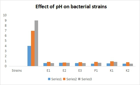
Figure 2. Effect of Varying pH on Bacterial Growth
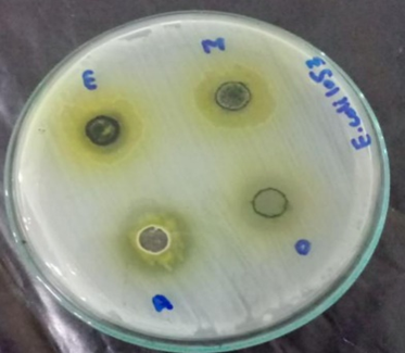
Figure 3. Zones of Inhibition of M. oleifera Extracts against Isolated Bacterial Strains
Table 3. Antibacterial Activity of Moringa oleifera Extracts
|
Bacterial strains |
Zones of inhibition (mm) |
|||
|
Ethanol |
Methanol |
DMSO |
Aqueous |
|
|
E1 |
17 mm |
24 mm |
15 mm |
23 mm |
|
E2 |
22 mm |
20 mm |
19 mm |
25 mm |
|
E3 |
15 mm |
10 mm |
0 mm |
20 mm |
|
P1 |
16 mm |
18 mm |
17 mm |
15 mm |
|
K1 |
22 mm |
21 mm |
10 mm |
0 mm |
|
K2 |
11 mm |
10 mm |
12 mm |
0 mm |
3.4. Disc Diffusion Assay
After incubation, inhibitory effects of antibiotic discs were observed in the zones of inhibition (millimeters) (Table 4). All organisms showed distinct activities against the selected antibiotics. All bacterial strains were sensitive against amikacin. Further, all bacterial strains were resistant against erythromycin, clindamycin, ceftriaxone, and cloxacillin. E3 strain was sensitive against ampicillin, penicillin, and amoxicillin. E3 and K2 strains were sensitive against tetracycline. K1, K2, and P1 bacterial strains were more sensitive against levofloxin and ciprofloxacin. E3 bacterial strain was highly sensitive against fosfomycin (Table 4).
Table 4. Zones of Inhibition of Antibiotic Discs against Pathogenic Bacterial Isolates
|
Zone of inhibition (mm) |
Pathogenic strains |
||||||
|
Antibiotics |
E1 |
E2 |
E3 |
P1 |
K1 |
K2 |
|
|
Penicillin |
R |
R |
S |
R |
R |
R |
|
|
Tetracycline |
R |
R |
S |
R |
R |
S |
|
|
Erythromycin |
R |
R |
R |
R |
R |
R |
|
|
Ampicillin |
R |
R |
S |
R |
R |
R |
|
|
Amoxicillin |
R |
R |
S |
R |
R |
R |
|
|
Ceftriaxone |
R |
R |
R |
R |
R |
R |
|
|
Clindamycin |
R |
R |
R |
R |
R |
R |
|
|
Fosfomycin |
S |
S |
S |
R |
S |
S |
|
|
Ciprofloxacin |
R |
R |
R |
S |
S |
S |
|
|
Levofloxin |
R |
R |
R |
S |
S |
S |
|
|
Amikacin |
S |
S |
S |
S |
S |
S |
|
|
Cloxacillin |
R |
R |
R |
R |
R |
R |
|
|
E1 |
Legends: R = Resistant is <12 mm, S = Sensitive is >15 mm
3.5. Biofilm Formation
The ability of biofilm formation was observed by using the following assays, that is, ring test and slime production.
3.6. Qualitative Ring Test
The formation of biofilms was examined qualitatively via rings formation stained with crystal violet. The potential for biofilm formation was judged subsequently after 24, 48, and 72 h of incubation. Biofilms adhere to the surface of test tubes by organizing rings. In the current study, these rings were stained by 0.5% crystal violet. It was found that the potency of bacterial strains attaching to the surface of test tubes was weaker after 24 h of incubation than after 48 h. Due to the staining of biofilm rings by crystal violet, the thickness of rings was strong and the color was dark after 72 h. It was also noted that with the addition of ethanol, methanol, DMSO, and aqueous extracts of M. oleifera, the potency of biofilm formation became relatively weak. Ethanol and methanol extracts manifested greater effects of inhibition on biofilm formation after 72 h (Figure 4). Whereas, the inhibitory effect of water and DMSO extracts on biofilm formation was lesser as compared to that of ethanol and methanol.
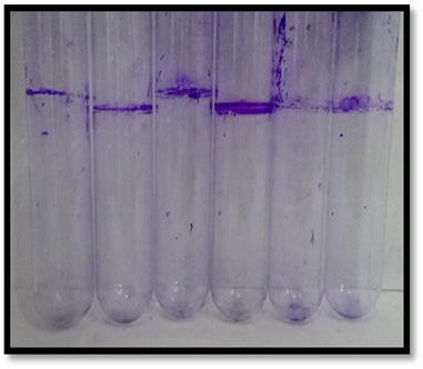
Figure 4. Bacterial biofilm rings stained by crystal violet
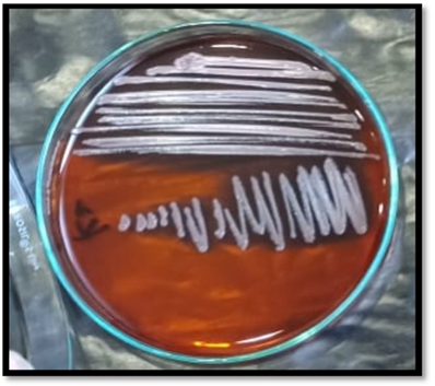
Figure 5. A) Bacteria on CRA Medium without Glucose, B) Bacteria on CRA Medium with Glucose
3.7. Slime Production
Congo red agar medium was prepared with and without glucose. All strains were cultured on CRA plate and incubated at 37°C for 24 h. Resultantly, changes of color were observed. Slime producing organisms showed dark black color on CRA plates. All strains produced dark black colonies on CRA plates, except P1 with glucose. All strains showed pink color on CRA medium without glucose (Figure 5). E2 and E3 are described as powerful creators of slime. However, in the case of Congo red agar test, no difference was observed in the effect of solvents on bacterial strains.
MDR bacterial infections are one of the major threats to global health. They are among the main reasons of increased morbidity and mortality in the world [24]. The reason of drug instability is the repeated use and misuse of antibiotics, as well as the lack of development of new drugs due to few economic incentives and difficult regulatory requirements. It is well-known that due to the development of host biofilms diseases become generally difficult to cure, which aids the spread of microbes and serious infections [25]. Therefore, there is an intense need to produce new antimicrobial drugs that may prevent the evolution or cause the elimination of whole biofilms. Antimicrobials derived from plants have been proved to be one of the most propitious sources observed as safe because of their native origin, as compared to manufactured compounds [26].
In this research, bacterial strains were isolated from urine, blood, and sputum samples to investigate their biofilm forming capacity. After conducting literature review on many medicinal plants, M. oleifera was selected and the leaves extract of this plant were used to check their affectivity against previously isolated pathogens. The isolated strains were labeled as E1, E2, E3, K1, K2, and P1. These strains were distinguished on the bases of their color, size, appearance, margins, elevations, and opacity. Different types of biochemical tests were performed for further differentiation. After conducting these tests, these bacterial strains were identified as Gram negative rods with qualities similar to the genus Escherichia coli, Pseudomonas, and Klebsiella. The variations discovered in the current research are related to other variations discovered by Paudyal et al. [27].
Secondly, physiological parameters were tested on the strains, such as the effect of varying temperature and pH. It was observed that all organisms grow best at temperature 37 °C and pH 7. Higher temperature and pH have an adverse association with the increase in bacterial growth. Bar graphs were drawn using Excel and these graphs showed significant differences. Another study reported that pathogenic bacteria grow at 3-10 pH values and 4-90°C temperature. Bacterial growth was approximated by the demonstration of their growth curve in this study [28]. All organisms went through a latency phase in the first 2 h to prepare themselves for division. Then, cells entered into the log phase that extended almost 4 to 24 h in all strains. Bacterial recognition was conducted through genetic analysis by using 16S rRNA sequencing. Due to its high accuracy and efficiency, 16S rRNA target sequencing technology has quickly become the most widely used approach to describe microorganisms [29].
oleifera leaves extract was prepared in DMSO, methanol, ethanol, and water and their affectivity against pathogens was observed by using agar well diffusion method. It was observed that some strains showed inhibitory effects (25 mm) in aqueous extracts. Most of them showed better inhibitory effects through ethanolic and methanolic extracts of M. oleifera leaves. Ethanol and methanol are the most suitable for plant extraction because of their apposed nature that ensures the release of a wide range of biologically active extracts from plants [30]. Antibacterial effects of antibiotics, such as penicillin, tetracycline, erythromycin, ampicillin, amoxicillin, ceftriaxone, clindamycin, fosfomycin, ciprofloxacin, levofloxacin, amikacin, and cloxacillin were checked by using disc diffusion assay. All bacterial strains were sensitive against amikacin. E3 bacterial strain was highly sensitive against fosfomycin. Another study reported amikacin as highly effective against all bacterial strains and ampicillin as the least effective [31].
The biofilm forming ability of organisms was checked by using ring test for 24 hours to 72 hours [32]. The results were unexpected because all strains produced biofilm which became thicker and thicker as time proceeded. This was seen through crystal violet staining. After 72 h, rings formed around the walls of test tubes. This test was quantitatively performed in 96 well microtiter plate and the readings were noted. The same pattern of biofilm initiation, maturity, and distance was observed [33]. Slime production test was also performed on Congo red agar. The slime producing bacteria showed black color growth on the medium. All organisms produced dark black colonies on CRA plate, except P1 with glucose. All strains observed pink colonies on CRA medium without glucose. E2 and E3 became visible as strong slime producers.
4.1. Conclusion
The study concludes that M. oleifera leaves extracts are highly effective against pathogenic bacteria. They have antibacterial and antibiofilm characteristics. Therefore, detailed extraction and characterization of these extracts may lead us towards the development of an effective and economical solution to antimicrobial drug resistance. To achieve this goal, extensive research should be carried out on the structural elucidation of bioactive compounds in M. oleifera. Further, in vivo studies should be carried out to completely investigate the mechanism of action of M. oleifera extracts against bacterial infections.