Genetic Variability of Xanthomonas citri pv. citri in South Punjab, Pakistan
Sufyan Raza1, Hasan Riaz1*, Faheem Zia1, Seema Kanwal1, Sabeeh Khan1, Muhammad Hassan1, Sami Ullah2, Mirza Abdul Qayyum1, Nadeem Ahmed1, Muhammad Imran3, Muhammad Shahzad Zafar4, Maryam Zain5
1Institute of Plant Protection, MNS University of Agriculture Multan
2Department of Horticulture, MNS University of Agriculture Multan
3Department of Soil and Environmental Sciences, MNS University of Agriculture Multan
4Mango Research Institute, Multan
5Department of Biochemistry and Biotechnology, The Women University Multan
Abstract
Background. Citrus is an important nutritious fruit that belongs to the family Rutaceae, comprising a large group of trees and shrubs. Citrus constitutes about 40% of all fruits grown in Pakistan. The total contribution of kinnow, grown in Pakistan, in the international market is US$ 222 million. In Pakistan, citrus production has been recorded up to 2.0 million metric tons. There are multiple abiotic and biotic factors involved in the low productivity of citrus in the country. Moreover, citrus canker (caused by the pathogen Xanthomonas citri pv. citri) has been the major threat for citrus production in Pakistan for the last 10 years. The current study aims to document the disease incidence of citrus canker along with the characterization of citrus canker causal organism.
Method. Samples exhibiting disease symptoms (such as small pimple-like spots about 1 mm in diameter and yellow in colour) were collected from the target areas. After isolation and purification, the samples were subjected to PCR analysis for molecular identification and characterization through SDT and MEGA11 software packages.
Results. A total of 112 samples were collected from different sites in South Punjab, Pakistan. The bacterial colonies were small and yellow in color, which turned pink after staining. Bubble formation was also observed after conducting biochemical tests. Genetic analysis showed that Xanthomonas citri pv. citri Multan isolate has 98.91% nucleotide identity with Indian, Korean, Chinese, and South African isolates.
Conclusion. The study underlines the emerging pathogen population of citrus canker that could help to manage this disease.
Highlights
- Citrus canker is an emerging threat in South Punjab, Pakistan.
- Citrus canker has high incidence as compared to previous reports.
- The prevailing citrus canker pathogen is an isolate Xanthomonas citri citri reported from various regions of the world.
1. Introduction
Citrus is the world's most valuable commercial and nutritional commodity, belonging to the Rutaceae family. It is also Pakistan's most important fruit crop. Citrus fruits are nutrient-dense, juicy, and delicious. Citrus limon, Citrus sinensis, mandarin, Citrus aurantifolia (lime), and Citrus paradise (grapefruit) are all members of the citrus genus [1].
Citrus is a tropical and subtropical fruit family that originated in China, India, Indonesia, and Malaysia. It is now produced all over the world, especially in tropical and subtropical climes. Citrus production accounts for 30% of the total fruit production in Pakistan [2]. The province of Punjab produced 97.1% of the country’s total citrus output (kinnow) in 2014-2015, while Khyber Pakhtunkhwa (KPK) produced 1.29%, Sindh produced 1.26%, and Baluchistan produced 0.29% [3]. Among all the citrus cultivars grown in Pakistan, the most important are mandarins (kinnow and feutrell) and sweet oranges [4]. Citrus is grown on 6.4 million hectares worldwide, with an annual production of 106.7 million tonnes. In Pakistan, citrus occupies 193.7 thousand acres with production up to 2.4 million tonnes, with an average per hectare yield of 12.4 tonnes [5].
Citrus canker disease is caused by the pathogen Xanthomonas citri pv. citri, which is the most destructive and threatening pathogen for citrus across the globe [6, 7]. Fruits, leaves, and twigs show the symptoms in case of citrus canker disease. The lesions initially appear as small, round, elevated patches that are bright green in colour [8]. Necrotic sores appear on leaves, fruits and branches as signs of pathogen infection. Lesions on stems, leaves, crowns, and fruit appear as brownish yellow, circular in shape, and 2-10 mm in size [9].
Genetic diversity is key to biological diversity and a prerequisite for the development of new organisms. In contrast to environmentally generated differences, which usually result in only transient, non-heritable modifications of the phenotype, genetic variability is marked by the production of genotypically different individuals or attested by the presence of different individuals [10].
Reunion and the nearby Indian Ocean islands are known to have a typical occurrence of the A strain of Xanthomonas citri. It has a high level of antibiotic resistance. A single strain known as "canker" was found in Mexico in 1981 on Mexican lime. Citrus fruits are widely grown in the South Punjab region of Pakistan. Citrus canker, caused by Xanthomonas citri, poses a grave threat to citrus in this region. In order to control citrus canker, prior knowledge of the bacterial population involved in inducing it is essential. Therefore, the current study is aimed at assessing genetic variability in Xc population and its phylogenetic relationship with the already reported Xc population from all over the world.
2. MATERIALS AND METHODS
2.1. Sample Collection, Isolation, and Purification
Several citrus fields in the South Punjab region were visited to collect disease samples from different locations including Shujabad, Muzaffargarh, Khanewal, Lodhran, and Vehari. Citrus canker disease was identified in various orchards and leaf samples were collected. Disease incidence was recorded on the basis of disease symptoms. Infected samples were collected and placed in sterile polythene bags for further analysis. All samples were brought to the MNS-UAM diagnostic lab. Firstly, all samples were washed with tap water to remove soil particles and debris. Then, proper sterilization was done with 3% sodium hypochlorite for 2-3 minutes.
2.2. Morphological and Biochemical Characteristics of Xanthomonas citri pv. citri
Morphological identification of the pathogen was done on the basis of colony color and shape, observed with the naked eye and also under the microscope. For the biochemical confirmation of Xanthomonas citri pv. citri, KOH test, catalase test, and Gram staining were performed, as per the prescribed method [11–14].
2.3 Molecular Characterization of Citrus Canker Pathogen
2.3.1. Polymerase Chain Reaction (PCR) . DNA was isolated with the help of phenol chloroform method and quantification was done by nanodrop. After DNA extraction, the samples were subjected to PCR amplification by using the primers 27F (5′- AGAGTTTGATCCTGGCTC-3′) and 1391R (5′- GACGGCGGTGTGTRCA-3′) in a thermal cycler. Then, the 16S rRNA gene was amplified from a single colony of the culture (Nyx, Technic, Inc., USA). DNA template, 15 μL of nuclease-free ddH2O, 10μM each of forward and reverse primers, 1.5mM of MgCl2, 1X each of Taq buffer, and 5U Taq polymerase (Takara, Japan) were used in a 25ml PCR reaction.
2.3.2. Sequencing and Phylogenetic Analysis. After the sequencing of the PCR product, the obtained sequence was subjected to BLAST analysis to confirm its nucleotide identity. The alignment of all the retrieved sequences was done with the help of MUSCLE alignment tool embedded in Sequence Demarcation Tool (SDT). Phylogenetic tree was constructed by using MEGA11 software package [15, 16].
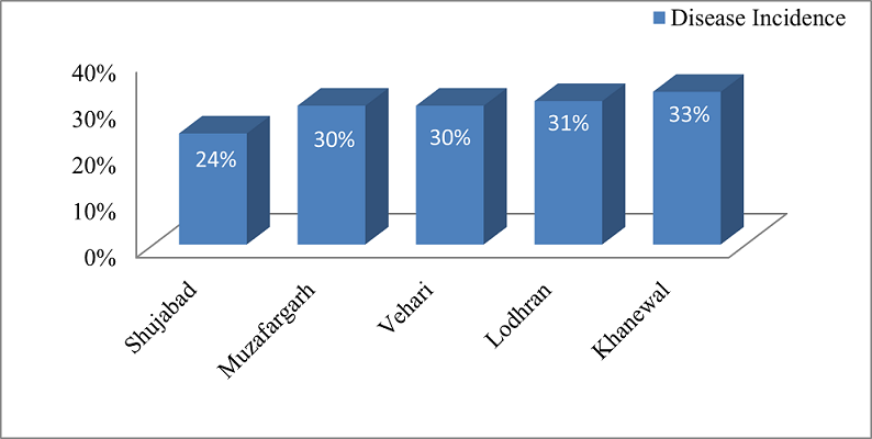
Figure 1. Disease Incidence of Citrus Canker
3. RESULTS
3.1. Disease Incidence
Infected samples with symptoms such as warty, rust-brown to tan spots, rough to the touch, and surrounded by a yellow halo were collected from the fields and brought to the laboratory for the isolation and identification of the pathogen. The disease incidence date was also recorded during survey and sample collection on the basis of symptomology. The maximum disease incidence was recorded in Khanewal at 33%, followed by Lodhran at 31%, and Muzafargarh and Vehari at 30%, respectively. Whereas, the minimum disease incidence was recorded in Shujabad at 24% (Figure 1).
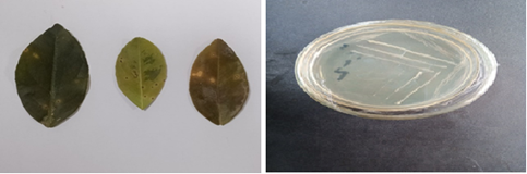
Figure 2. Infected Samples Showing Citrus Canker Symptoms and Isolated Bacterium on NA Media
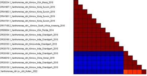
Figure 3. Similarity Identity Matrix Based on Pairwise Sequence Comparison of Xanthomonas citri pv. citri
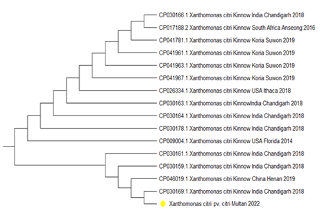
Figure 4. Phylogenetic Analysis of Xanthomonas axanopodis pv.citri Multan Isolate 2022
3.2. Morphological and Biochemical Characteristics of Xanthomonas citri pv. citri
The colony color of the bacterial isolate was light yellow to dark yellow and the colonies were round and small in size (Figure 2). Gram staining showed that the isolated bacteria were gram-negative and appeared as rod shaped under the microscope. In case of biochemical tests, bacterial isolates were positive in hydrogen peroxide production, as well as in catalase production and KOH production.
3.3. Molecular Characterization
Polymerase chain reaction (PCR) amplified approximately 1400bp band of 16s rRNA gene sequence from the collected isolates of Xanthomonas citri pv. citri. The amplified products were sequenced at Macrogen Korea. The BLAST analysis, based on 16s RNA gene sequence, confirmed the bacterial sequence to be an isolate of Xanthomonas citri pv. citri. The pairwise sequence comparison revealed 99.19% homology of Multan 2022 isolate with Xanthomonas citri pv. citri isolates from India, China, USA, Korea, and South Africa, with accession numbers CP030169, CP030166, CP030159, CP030161, and CP046019, respectively (Figure 3). The phylogenetic analysis clustered the Multan isolate 2022 with Chinese and Indian isolates (Figure 4).
4. DISCUSSION
Citrus is a prominent member of the Rutaceae family due to its appetizing flavor and appealing appearance. Citrus fruits are primarily processed for juice production in the beverage industry and also used for various dishes [17]. In this research, it was observed that Xanthomonas citri pv. citri has a colony color which varies between light yellow and dark yellow. Previous studies also revealed that this bacteria is gram-negative and reflects yellow colony color on the agar plate. In previous studies, yellow pigmented and mucoid colonies were formed on agar media plates due to the pigmentation of Xanthomonas [18]. In case of biochemical tests, bacterial isolates were positive in hydrogen peroxide production, as well as in catalase production and KOH production. This is in line with the results of a previous study conducted in India in which Xc from the Marathwada region of India showed similar characteristics [19]. The molecular analysis, similarity matrix, and phylogeny showed that the isolate Multan 2022 has maximum resemblance with the isolates of India, China, USA, Korea, and South Africa. The previously reported studies also prescribed that Xanthomonas citri pv. citri isolated from India has 100% nucleotide similarity with the isolates of USA and China [20, 21].
4.1 Conclusion
Citrus fruits are widely grown in the South Punjab region and are heavily infected with Xanthomonas citri pv. citri, the causal organism of citrus canker, which poses a grave threat to citrus in the said region. The current study gave a vivid picture of citrus canker disease incidence in South Punjab, as well as the species and strains responsible for inducing citrus canker in the region. Based on the study, it is imperative to comb the area for citrus canker and adopt management measures to limit its spread by introducing disease free nursery, treatment with appropriate bactericides, and eradicating the heavily infected citrus orchards.
Conflict of Interest
The author of the manuscript has no financial or non-financial conflict of interest in the subject matter or materials discussed in this manuscript.
Data Availability Statement
The data associated with this study will be provided by the corresponding author upon request.
Funding details
This research did not receive grant from any funding source or agency.
Bibliography
- Awan MZ, Ishfaq M, Hafiz IA, Ijaz M, Ayyub CM. Incidence of citrus canker in Barani area at Chakwal. Presented at: Proceedings of the 1st International Seminar on Citrus; December 2–5, 1992; Pakistan.
- Siddique MI, Garnevska E. Citrus value chain: a survey of Pakistan citrus industry. In: Egilmez G, ed. Agric Value Chain. BOD; 2018:37–56. https://doi.org/10.5772/intechopen.70161
- Mal B, Singh SS, Ghodake R. Investment in agricultural research for sustainable development in Asia and the pacific. Food and Agriculture Organization of Pakistan. https://www.apaari.org/web/wp-content/uploads/2017/Country%20Status%20Report_29-7-2017_2.pdf
- Ayub M, Jahangir HS, Mumtaz K, Amin M. Pathogenic variation and host range of campestris pv. citri isolates. Pak J Phytopathol. 1996:e18.
- Ference C, Gochez A, Behlau F, Wang N, Graham J, Jones JB. Recent advances in the understanding of Xanthomonas citri ssp. citri pathogenesis and citrus canker disease management. Mol Plant Pathol. 2018;19:1302–1318. https://doi.org /10.1111/mpp.12638
- Schaad NW, Postnikova E, Lacy G, et al. Emended classification of Xanthomonas pathogens on citrus. Papers in Plant Pathol. 2006:e96. https://doi.org/10.1016/j.syapm.2006.08.001
- Ali S, Hameed A, Muhae-Ud-Din G, Ikhlaq M, Ashfaq M, Atiq M, Ali F, Zia ZU, Naqvi SAH, Wang Y. Citrus Canker: A Persistent Threat to the Worldwide Citrus Industry—An Analysis. 2023;13:1112. https://doi.org/10.3390/agronomy13041112.
- Izadiyan M, Taghavi SM, Farahbakhsh F. Characterization of Xanthomonas citri citri isolated from grapefruit in Iran. J Plant Pathol. 2018;100:257–267. https://doi.org/10. 1007/s42161-018-0081-3
- Stall RE, Seymour CP. Canker, a threat to citrus in the Gulf Coast states. Plant Dis. 1983;67(5):581–585.
- Tomilova OG, Shternshis MV. The effect of a preparation from Chaetomium fungi on the growth of phytopathogenic fungi. Appl Biochem Microbiol. 2002;42:67–71. https://doi. org/10.1134/S0003683806010108
- Bergey SA. Bergey’s Manual of Determinative Bacteriology. 9th ed. Philadelphia, PA: Williams and Wilkins; 1984.
- Iqbal MN, Anjum AA, Ali MA, et al. Assessment of microbial load of un-pasteurized fruit juices and in vitro antibacterial potential of honey against bacterial isolates. Open Microbiol J. 2015;9:26–31. https://doi.org/10.2174/ 1874285801509010026
- Braithwaite J. Setting standards for restorative justice. Br J Criminol. 2002;42:563–577. https://doi.org/10. 1093/bjc/42.3.563
- Al-Dulaimi F, AL-Kaisse AA, Al-Rubaye L, Abdulwadood M. First report of citrus bacterial canker caused by Xanthomonas citri citri in Iraq. Res J Biotechnol. 2018;12:24–31. http://dx.doi.org/10.24126/jobrc.2018.12.2.534
- Ausbel FH, Brent R, Kingston RE, Moore DD. Current Protocols in Molecular Biology. 4th ed. Canada: Wiley; 1994.
- Tamyra K. Akamatsu H, Asami K, Habitat conditions of Anaphalis margaritacea yedoensis (Franch. et Savat.) Kitam. on Asakura area of Maruyamagawa River. Humans Nat. 2007;18:45–49.
- Gottwald TR, Sun X, Riley T, Graham JH, Ferrandino F, Taylor EL. Georeferenced spatiotemporal analysis of the urban citrus canker epidemic in Florida. Phytopathology. 2002;92:361–377. https://doi.org/10. 1094/phyto.2002.92.4.361
- Das AK. Citrus canker-a review. J Appl Horti.2003;5(1):52–60.
- Mohammadi M, Mirzaee MR, Rahimian H. Physiological and biochemical characteristics of Iranian strains of Xanthomonas citri citri, the causal agent of citrus bacterial canker disease. J Phytopathol. 2001;149(2):65–75. https://doi.org/10. 1046/j.1439-0434.2001.00570.x
- Shehzadi I, Naz S. Morphological, biochemical and genetic characterization of citrus canker pathogen (Xanthomonas citri) from citrus cultivars of punjab, pakistan. J Ani Plant Sci. 2019: 29(1):117–124
- Park DS, Hyun JW, Park YJ, et al. Sensitive and specific detection of Xanthomonas citri pv. citri by PCR using pathovar specific primers based on hrpW gene sequences. Microbiol Res. 2006;161:145–149. https://doi. org/10.1016/j.micres.2005.07.005