
| Review | Open Access |
|---|
Veterinary Reproductive Ultrasonography: Advances, Applications, and Future Prospects |

|
|---|
![]() Sajad Ali Laghari* ,
Sajad Ali Laghari* ,
![]() Qudratullah Kalwar,
Qudratullah Kalwar,
![]() Muhammad Mohsen Rahimoon,
Muhammad Mohsen Rahimoon,
![]() Fayaz Hussain,
Fayaz Hussain,
![]() Fazul U Rahman Soomro,
Fazul U Rahman Soomro,
![]() Taj Muhammad,
Taj Muhammad,
![]() Abdul Saboor,
Abdul Saboor,
![]() Iqrar Uddin
Iqrar Uddin
1Department of Theriogenology, Faculty of Veterinary Sciences, Shaheed Benazir Bhutto University of Veterinary and Animal Sciences, Sakran, Pakistan
2Department of Wildlife Management, Shaheed Benazir Bhutto University of Veterinary and Animal Sciences Sakrand Pakistan
Background. Ultrasonography is a vital imaging tool in veterinary practice, particularly for reproductive assessment and pregnancy monitoring in animals. Modern ultrasound devices are compact, affordable, and efficient, facilitating their widespread clinical use. Initially applied in livestock for early pregnancy detection in the 1980s, this technique now aids in managing reproductive disorders. Despite its benefits, only a limited number of veterinarians utilize it beyond basic pregnancy diagnosis.
Methods. This review examines the current literature on veterinary reproductive ultrasonography, as well as its various applications, types, modes including A-mode (1D), B-mode (2D), M-mode (motion), and Doppler (blood flow), diagnostic purposes, proper probe selection, patient preparation (hair trimming, gel application), and machine settings. Data from several studies are analyzed to summarize the uses of reproductive ultrasonography in domestic animals.
Results. Ultrasonography enables early pregnancy detection (as early as Day 20-25 in small ruminants and Day 23-30 in cattle), fetal viability assessment, and sex determination (Day 55-60). It accurately monitors ovarian structures (follicles, corpus luteum) and detects reproductive disorders (endometritis, pyometra). Doppler ultrasound evaluates blood flow, while M-mode tracks fetal heart activity. This technique is safe, non-invasive, and enhances the efficiency of reproductive management.
Conclusion. Ultrasonography is a safe, non-invasive, and a highly effective tool for reproductive management in veterinary medicine. It enhances early pregnancy diagnosis, fetal monitoring, and infertility management in livestock and companion animals. Despite its advantages, underutilization persists due to limited expertise among practitioners. Further research is needed to explore its long-term biological effects, though current evidence supports its diagnostic safety and reliability.
Highlights
- Non-invasive, real-time imaging for reproductive assessment in animals.
- Early pregnancy detection (Day 20-30) and fetal viability monitoring.
- Doppler and M-mode ultrasound assess blood flow and cardiac activity.
- Safe, cost-effective, but requires skilled operators for accurate diagnosis.
GRAPHICAL ABSTRACT

1. INTRODUCTION
Ultrasonography is a widely utilized imaging technique in veterinary practice [1]. It is the preferred imaging technique for assessing the reproductive system and monitoring pregnancy in both animals and humans. Modern ultrasound devices are now more compact, affordable, and highly efficient. These advancements, combined with the use of digital imaging technology, have facilitated the widespread use of ultrasound in clinical practice environments [2–4]. During the 1980s, /ultrasonography was first applied for early pregnancy detection in livestock. Various ultrasonographic modalities, including A-mode, B-mode, and Doppler mode, have been employed in the livestock sector for reproductive assessment. Ultrasonography has many applications in reproductive management of livestock and companion animals. However, it still remains underused by veterinarians for managing reproductive disorders [5]. Additionally, many veterinary ultrasonographers primarily rely on ultrasound scanners for early pregnancy detection and research. The routine use of ultrasonography enhances diagnostic accuracy, leading to more precise drug administration. In large ruminants, it significantly reduces or even eliminates the common errors associated with manual rectal palpation when assessing physiological and pathological conditions of the ovaries as well as the uterus [6].
Real-time B-mode diagnostic ultrasound has been gaining popularity as a useful imaging technique for small ruminant reproduction. Its effectiveness in addressing unresolved issues, diagnosing pregnancy, and managing reproductive disorders has become increasingly evident [7]. No cases of embryonic mortality or abortion have been observed, as continuous ultrasound monitoring throughout pregnancy confirmed that the newborns were anatomically normal and healthy [8]. Ultrasonography is widely utilized in veterinary gynecology to determine pregnancy stages and count fetuses. The trans rectal method allows for precise diagnosis as early as day 23 of gestation [9, 10], while the trans-abdominal method becomes effective by Day 35 of the pregnancy [11].
Reproductive ultrasonography has greatly advanced in diagnosing infertility in dogs [12, 13]. In the past, its application in canine reproduction was primarily limited to pregnancy detection. However, with technological advancements, its use has expanded beyond diagnosis to include pregnancy monitoring, estimation of parturition dates, and management of various conditions such as threatened or induced abortions, endometritis, pyometra, subinvolution, pyometra, and ovarian disorders [14]. Today, ultrasonography serves not only as a diagnostic method but also as a means to monitor the effectiveness of treatments for various reproductive disorders. Hence, it is evident that veterinary reproductive ultrasonography is a valuable tool for controlling fertility in companion and farm animals.
1.1. Basic Principle of UltrasonographyThe basic principle of ultrasound relies on using high frequency sound waves to produce pictures of the internal organs. These sound waves are produced by a transducer. They pass through tissues and return when they come into contact with varying densities. When the transducer detects the returning echoes, it transforms them into electrical signals. A computer then processes these signals to create a real-time image. This technique is commonly used in medical and veterinary diagnostics to visualize organs, tissues, and developing fetuses without using radiation [15]. It is important to understand that the ultrasound image represents a thin slice of an organ, resembling a low-magnification histology section. The probe essentially acts as a blade, cutting through the organ from top to bottom. As a result, a two-dimensional, cross-sectional image of the targeted organ is produced using B-mode ultrasonography. As the probe glides over the tissue, ultrasound images are continuously refreshed and layered on top of one another. This rapid succession of sectional views creates an effect similar to animation, making the structures appear to be in motion. A clear understanding of the organ’s three-dimensional form in space is essential to accurately interpret the sectional images displayed on screen [6].
1.2. Properties and Image Clarity of Probes of UltrasoundThe most sensitive part of the ultrasonic system is the probe. In veterinary practice, probes with frequencies ranging from 3 to 10 MHz are commonly used. There are three main types of probes: linear, curvilinear, and sector. In theriogenology, linear or curvilinear probes are typically used for transrectal ultrasound examination of the ovaries and uterus. The linear probe contains a fixed arrangement of electronic crystals activated to produce a rectangular sonograph [16]. This probe provides high-resolution imaging for tissues located near the surface. In contrast, curvilinear and sector probes have one or more crystals arranged to produce a pie-shaped ultrasound beam. A key advantage of these probes is their ability to scan a larger area without needing extensive contact with the surface. When using IVF technology, the sector probe is especially helpful for ultrasound-guided transvaginal aspiration of bovine follicles and for fetal inspection during transabdominal ultrasonography. Lower-frequency probes allow for deeper tissue penetration but result in reduced image resolution [17].
Table 1. Probe Selection Guidelines for Reproductive Ultrasonography in Different Animals
|
Species |
Probe Type |
Frequency Range (MHz) |
Depth Penetration |
References |
|---|---|---|---|---|
|
Cattle/Buffalo |
Linear/Curvilinear |
5–10 MHz |
5–12 cm |
[9, 16] |
|
Goat/Sheep |
Microconvex/Linear |
6–8 MHz |
4–8 cm |
[11] |
|
Horse |
Linear (transrectal) |
5–7.5 MHz |
8–15 cm |
[9. 16, 58] |
|
Dogs |
Curvilinear/Sector |
3–6 MHz |
5–15 cm |
[27] |
|
Cats |
High-frequency Linear |
7.5–10 MHz |
2–6 cm |
[27] |
1.3.1. Amplitude Mode (A-mode). Amplitude mode, also known as 1-D ultrasound, is the earliest ultrasound technique, developed in 1930. In this method, the transducer emits a single ultrasound pulse into the medium, producing a basic one-dimensional image. When the ultrasound waves reach tissue boundaries, they generate a series of vertical peaks on the display. To find the distance between these echo spikes, the speed of sound in tissue (1540 m/s) is divided by half the measured time for the echoes to return. However, this technique offers minimal information about the spatial relationships of the imaged structures. Amplitude mode (A-mode) ultrasound produces a one-dimensional representation that displays echo amplitudes at different depths. This mode utilizes amplitude and depth as its primary axes. It is commonly employed for pregnancy diagnosis in goats, sheep, and equine [18].
1.3.2. Brightness Mode (B-mode). B-mode ultrasonography produces a two-dimensional (2D) image by scanning an area with a linear array of 100 to 300 piezoelectric elements. This is unlike A-mode, which utilizes a single element. B-mode ultrasound, commonly referred to as Brightness-mode imaging, displays two-dimensional anatomical structures with varying brightness to represent echo amplitudes. Grayscale intensity indicates echo strength, while the horizontal and vertical axes show actual tissue distances. This technique allows for the visualization of a cross-sectional image of the targeted area. In the majority of modern ultrasound systems, B-mode is used to examine the reproductive tract in animals using real-time scanning. This imaging technique provides a two-dimensional display where brightness corresponds to echo amplitude. Real-time imaging continuously captures echoes, allowing dynamic observation of events, such as fetal limb movements and heartbeat in real-time [19].
1.3.3. Motion Mode (M-mode). B-mode imaging has been modified to create M-mode (motion mode) imaging that evaluates dynamic structures like the heart. It represents changes in reflector depth over time using a basic line graph, where depth and time serve as the axes [6]. When an A-mode or B-mode image is Trasferred in M-mode ultrasonography, pulses are sent out quickly. This process is similar to creating a video in ultrasound. As reflective organ boundaries shift in relation to the probe, this technique helps measure the velocity of specific organ structures, effectively illustrating their movement over time. Firstly, a two-dimensional image is obtained and a single scan line is positioned over the area of interest. M-mode then displays how the structures along that line shift toward or away from the probe over time. With its high temporal resolution, M-mode is particularly effective for/in identifying and capturing fast movements [20].
1.3.4. Doppler Mode. Doppler ultrasound systems create vibrant, multicolored images of blood flow patterns by detecting its movement at an angle to, away from, or in the direction of the transducer. Doppler Shift refers to the alteration in frequency or wavelength of a sound wave due to the relative motion between the sound source and receiver. This principle helps estimate high-frequency ultrasonic waves off of moving red blood cells to reflect blood flow in arteries. Doppler ultrasound specifically detects blood flow, in contrast to normal ultrasonography, which uses sound waves to create images [21].
1.4. Ultrasonographic TechniquesIn buffaloes, horses, goats, sheep, and pigs, both transrectal and trans-abdominal ultrasound examinations are commonly used to assess reproductive health, including normal and abnormal conditions. During the middle to late stages of pregnancy, trans-abdominal ultrasonography is typically conducted while the animal remains standing to evaluate fetal well-being by monitoring movements, heartbeats, umbilical blood flow, and the condition of the placentomes. The quality of the ultrasound image is influenced by the selected transducer, the machine's gain settings, and patient preparation. One step in the preparation of the patient for diagnosis is to trim the hair over the region of concern. Hair traps air, making it difficult for sound to travel. By moistening the hair with water or alcohol, air can be eliminated from areas where the hair is fine or thin. After the hair has been trimmed or wet, ultrasonographic gel is used to ensure the best possible contact and sound transmission from the transducer to the animal's tissues [22]. The machine's gain settings are used to adjust how loud the echo comes back from the structure of interest. The equipment can be adjusted to compensate for the loss of signal because the ultrasonic beam's power diminishes as it moves deeper into the tissues. With a slope that accounts for the growing signal loss brought on by sound reflection and refraction from tissues positioned between the transducer and the deepest structure to be scanned, this compensation often varies in machines. The thickness of the region being inspected should be taken into consideration while choosing the transducer, and if at all possible, the focused zone of the transducer should correspond to the depth of the overall area of interest [23].
1.4.1. Trans-rectal Ultrasound Technique in Cattle and Buffaloes. For trans-rectal ultrasound scanning, a linear or curved probe operating within a frequency spectrum of 5 to 10 MHz is commonly employed. The preparation process for this procedure is similar to that of rectal palpation. The transducer is coated with gel, although the natural moisture of the rectal wall often makes additional coupling medium unnecessary. To prevent rectal injuries, the same precautions taken during rectal palpation should be followed when inserting the lubricated hand and transducer. Additionally, fecal matter must be cleared beforehand to prevent image distortions during the examination. It is crucial to take appropriate precautions to ensure that the transducer is correctly guided through the rectal passage and avoids entering an unintended cavity. This can be accomplished by extending the fingers beyond the tip of the transducer during substantial forward movements. The trans-rectal technique is the preferred method for ultrasound examination in non-pregnant females and those in the early stages of pregnancy [24]. To secure either a linear or curvilinear probe AI sheaths or PVC pipes can be utilized, making it more convenient for trans-rectal scanning in small ruminants [10]. In small ruminants and swine, trans-rectal examination is typically performed while the animal is manually restrained in a standing position. To enhance image quality, feces should be cleared from the rectum. Moreover, a water-soluble gel should be applied beforehand to prevent mucosal injury and facilitate sound wave transmission. A probe operating at a frequency of 6 to 8 MHz is introduced into the rectum, aligning the transducer perpendicular to the ventral abdominal wall. The urinary bladder is the initial structure detected, presenting as an echogenic formation. The uterine body is located dorsal to the bladder, with each uterine horn positioned on its respective side. The exact placement of the uterus depends on factors such as bladder fullness, uterine size, the animal’s age, and reproductive history. After locating the bladder, the probe should be rotated laterally in both clockwise and counterclockwise directions to achieve a comprehensive view of the uterine horns and ovaries. The transrectal approach enables reliable pregnancy diagnosis in small ruminants and pigs, as early as 20 days post-breeding [25].
1.4.2. Trans-abdominal Ultrasonography in Farm Animals. In the middle to late stages of pregnancy, trans-abdominal ultrasonography is commonly conducted while the animal remains standing to assess fetal condition, including movements, heartbeats, integrity of the placenta, and umbilical blood flow. In both large and small ruminants, a convex transducer operating at a frequency of 3 to 5 MHz should be placed in the inguinal area, directly in front of the udder; in pigs, however, it should be placed just above the last teat [25]. To improve contact between the transducer and the skin while minimizing air interference, some breeds may have their inguinal hair clipped, while contact gel should be liberally applied to the probe. In large ruminants, the trans-abdominal approach is primarily used during mid to late gestation, as the fetus shifts into the abdominal area during this stage. However, Transrectal scanning is generally preferred for monitoring the fetus and other pregnancy-related aspects [26].
1.4.3. Trans-abdominal Ultrasonography in Companion Animals. A convex sector transducer operating at a frequency of 3 to 6 MHz is perfect for canines to assess the normal uterus, as it covers a larger area. While, in felines, high-frequency transducers of 7.5 to 10 MHz are preferred. Both species are typically examined in dorsal, lateral recumbency or while standing. However, many pet owners may resist shaving the ventral abdominal hair, which is a common practice to achieve optimal image clarity [27]. Using alcohol or similar wetting solutions before applying ultrasound gel on furry skin can improve imaging results by minimizing the interference caused by hair between the transducer and the skin. A distended urinary bladder enhances uterine visibility by serving as an acoustic window, positioning the uterine body between the bladder ventrally and the colon dorsally for clearer observation [28].
1.4.4. Ultrasound Applications in Reproductive Management of Domestic Animals. Ultrasound is better than radiography in examining soft tissue structures because it can distinguish between different soft tissues according to their composition and also separate fluid from them [29]. Therefore, ultrasound provides a non-invasive substitute for numerous radiographic contrast procedures, though both methods are viewed as complementary. Additionally, ultrasound offers data that was required exploratory laparotomy [30]. Other applications include pregnancy detection, determining fetal numbers, and even fetal sex [31]. As a method to diagnose pregnancy, transrectal ultrasonography is both precise and quick, providing immediate results at the time of testing. Factors such as the rate of embryonic loss and the effectiveness of rebreeding strategies at different stages after breeding also influence the benefits and drawbacks of when pregnancy diagnosis and resynchronization are performed [32].
1.5. Evaluation of Typical Ovarian Anatomical Components1.5.1. Follicles. The sonographic anatomy of bovine ovaries has been extensively studied. Antral follicles, which appear as anechoic (fluid-filled) regions, vary in size and can be distinguished from blood vessels in cross-sectional views due to the vessels’ elongated, tubular morphology [33]. Studies confirm a strong linear relationship between antral follicle diameters measured via transrectal ultrasonography in live animals and those obtained from postmortem examinations [34]. Studies have also recorded correlation coefficients ranging from 0.7 to 0.9 for different follicular sizes, when comparing in vivo ultrasonography with post-mortem examination of excised ovaries [35]. In goats, transrectal ultrasonography has been identified as a dependable technique for analyzing follicular dynamics [36].
1.5.2. Ovulation. Ultrasound examination has been utilized to identify ovulation, as documented in various studies [37]. A longitudinal study involving eight heifers employed ultrasonography to examine ovarian dynamics at 4-hour intervals throughout estrus and the subsequent period. The occurrence of ovulation was confirmed when a previously documented preovulatory follicle was no longer found to be detectable via ultrasonography, subsequently replaced by corpus luteum development at the identical ovarian site. The diagnostic accuracy of 2-hour interval ultrasound scanning to detect ovulation onset has been conclusively established [38].
1.5.3. Corpus Luteum. Multiple investigations have characterized the sonographic appearance of corpora lutea, documenting their distinctive morphological features [37, 39]. Typically, a corpora lutea can be identified via ultrasonography starting from the third day following ovulation. Ultrasonographic examination reveals developing corpora lutea as an irregular, ill-defined, greyish-black structure with echogenic spots inside the ovary. Mid-cycle corpora lutea exhibits characteristic ultrasonographic features including defined borders, homogeneous granular architecture, and moderate echogenicity that contrasts with the ovarian parenchyma. Luteal regression is characterized ultrasonographically by fading tissue demarcation, since the echogenic differential between the corpora lutea and ovarian parenchyma diminishes [40]. In small ruminants like goats, where rectal palpation cannot be used to assess ovarian structures, ultrasound imaging stands as the most effective method to monitor ovarian activity, as previously noted [36].
1.6. Ultrasonography Used in the Diagnosis of PregnancyEarly detection of pregnancy can enhance reproductive efficiency by shortening the time between consecutive artificial insemination procedures. Additionally, identifying non-pregnancy early on allows for the implementation of an assertive strategy to promptly rebreed the animal [41]. Pregnancy in cows can be detected through ultrasound imaging, which shows the fetus as a bright, reflective structure surrounded by a darker, fluid-filled space [42]. To manage early pregnancy losses, cows diagnosed as pregnant soon after breeding should receive additional check-ups in order to identify and rebreed those that suffer embryo loss. This guideline is relevant for all early pregnancy diagnosis techniques, such as transrectal palpation, conducted before the risk of embryonic loss decreases. Therefore, for optimal herd fertility, dairy producers using early pregnancy testing should strategically schedule follow-up checks to maximize reproductive efficiency.
1.6.1. Pregnancy Diagnosis in Cattle and Buffaloes. Early identification of non-pregnant animals post-breeding improves herd fertility outcomes in bovine species by reducing inter-service intervals and creating more opportunities for successful conception through timely re-insemination. Ultrasound provides a rapid method for pregnancy diagnosis. Skilled palpators can swiftly adapt to this technology by identifying the embryonic vesicle, developing embryo with a heartbeat, and corpus luteum verum amniotic membrane, as early as 30 days after breeding [31]. The stage of embryonic or fetal development can be assessed by taking crown-rump length (CRL) and transverse head diameter measurements at 30 days post-conception. Around this time, the CRL is approximately 1 cm and increases by about 1 mm per day until Day 40, after which the growth rate accelerates. In bovines (cattle and buffaloes), determining fetal age is the most clinically useful before Day 60 of pregnancy, as its diagnostic value diminishes considerably in later stages of gestation. The rationale for diagnosing pregnancy at 30 days in large ruminants is based on the 21-day estrus cycle; if an animal does not show signs of estrus 21 days after artificial insemination, ultrasonography can be used to examine it on Day 30 of gestation [43]. If the animal is confirmed pregnant on Day 30, it is beneficial; however, if it is not pregnant, estrus can be induced using either hormonal or non-hormonal protocols based on the condition of the ovaries and uterus. By implementing the 30-day pregnancy diagnosis approach, early identification of pregnancy is possible, along with the detection of any ovarian or uterine abnormalities. Additionally, this method allows for the precise use of fewer and more effective drugs for estrus induction, ultimately lowering treatment costs and reducing the time required for rebreeding [44].
1.6.2. Detection of Pregnancy in Goats and Sheep. Timely pregnancy detection in animals improves the reproductive efficiency and boosts pregnancy rates in small ruminants. By Day 25 post-breeding, experienced practitioners can effectively use ultrasound technology to detect the embryonic vesicle, a viable embryo with cardiac activity, distinct amniotic structures, umbilical attachments, and a functional corpus luteum. Furthermore, beginning at Day 25 post-breeding, embryonic/fetal development can be evaluated through biometric measurements of crown-rump length (CRL) and transverse head diameter (THD) [45] [30]. By Day 25, ultrasonographic examination also enables reliable identification of multiple pregnancies through simultaneous evaluation of distinct embryonic vesicles and corresponding corpora lutea bilaterally, which aids in managing pregnancy or delivery care. Beyond 50 days of pregnancy and into the later stages, trans-abdominal scanning is advised for optimal outcomes, as it minimizes the risks to pregnancy in small ruminants [11]. In the mid to late stages of gestation, the entire fetus cannot be fully visualized, making CRL measurement challenging. Instead, fetal age estimation is performed by evaluating the bi-parietal diameter and placentomes. For small ruminant herds with uncertain breeding histories, trans-abdominal ultrasonography serves as the recommended initial diagnostic approach. If no pregnancy-related structures are detected during this initial scan, transrectal scanning can then be performed to further confirm the reproductive status of the animal [6].
1.6.3. Pregnancy Diagnosis in Equine. Ultrasound has emerged as the ‘gold standard’ to assess a mare’s reproductive health. The application of grayscale ultrasound imaging has contributed to understanding key physiological aspects of mares, including their ovulation mechanisms, corpus luteum formation and development, follicular growth patterns, and embryonic migration dynamics [45–49]. It has been reported that Doppler ultrasonography technology allows for the evaluation of uterine and ovarian vascular dynamics in mares, as well as cyclic and early gestational changes [49, 50].
Transrectal ultrasound is frequently used to check for pregnancy in horses two to three (2-3) weeks after ovulation and for early embryo loss in horses five to six weeks (5-6) after ovulation. Regular ultrasonographic examinations afterwards are uncommon. Finding foals at higher risk of perinatal illness may also be facilitated by evaluating fetal development later in pregnancy [51]. In this regard, a low-frequency transducer (2–3.5 kHz) might be used to evaluate the horse fetus transabdominally during the latter two thirds of pregnancy [52, 53]. Previous studies utilized the measurements of fetal eye orbits, cranium, abdomen, thoracic width, intercostal spaces, and aorta to provide insights into fetal development in Thoroughbreds, Quarter Horses, Standardbreds, and Shetland Ponies between 4 and 11 months of gestation [51, 52, 54]. The best method for imaging the horse fetus in the early half of pregnancy is to use a convex transducer via the transrectal route. Furthermore, due to its structure, the rectum makes it very easy to change angle and gain cranial views; however, a linear transducer is limited to ventral views [53].
1.6.4. Detection of Pregnancy in Dogs and Cats. Uterine enlargement serves as an indicator of pregnancy, marked by the presence of a gestational sac. In dogs, these sacs are first detectable around day 20 post-breeding. For more accurate results, it is advisable to perform an ultrasonographic examination no earlier than Day 25 after the last breeding, as gestational sacs with viable embryos can be accurately detected by this stage. By Day 25 after breeding, the embryo adopts a semi-circular shape, the gestational sac develops an ampullary form, and the embryo’s heartbeat becomes detectable. Starting from Day 28, the gestational sac begins to elongate. Between Day 30 and 35, limb buds become visible. Finally, by Day 35-37, fetuses show notable movement with clearly detectable cardiac activity [27]. The gestational sac gradually becomes more elongated along the longitudinal axis. The mid-gestation period (Day 35-40) demonstrates sonographic evidence of organogenesis, with distinct visualization of the urinary and digestive systems concurrent with appendicular skeletal development. At this point, the fetal sac contains less fluid relative to the fetal mass, as compared to earlier stages. After 40 days, measuring the biparietal diameter (BPD) becomes straightforward due to the prominent ossification of bones. However, determining the CRL becomes challenging as the fetuses continue to grow. Additionally, bony structures like the ribs, vertebrae, and skull become clearly visible [55].
Table 2. Optimal Timing for Pregnancy Diagnosis Via Ultrasonography in Different Species
|
Species |
Earliest Detection (Days Post-Breeding) |
Preferred Method |
References |
|---|---|---|---|
|
Cattle |
23-30 days |
Transrectal |
[10, 31] |
|
Buffaloes |
25-30 days |
Transrectal |
[10, 31] |
|
Goat/Sheep |
20-25 days |
Transrectal |
[8; 11] |
|
Horse |
12-14 days |
Transrectal |
[9, 48, 54] |
|
Dogs |
20-25 days |
Transabdominal |
[27, 14] |
|
Cats |
15-20 days |
Transabdominal |
[27, 14] |
Real-time ultrasonography offers a distinct benefit as compared to other methods by enabling the identification of multiple fetuses [56]. Identifying the number of fetuses would enable producers to categorize animals carrying single, twin, or triplet pregnancies for tailored management practices. Bovine twin pregnancies can be accurately diagnosed via transrectal ultrasonographic evaluation during the day 40-55 gestational window, following artificial insemination [57]. Assessing fetal viability is a significant benefit of ultrasound as compared to other pregnancy diagnosis methods. During the examination, heart contractions can be observed between the ribs.
1.7.1. Ultrasonographic Identification of Fetal Sex. Determining the sex of a fetus holds significant importance in the animal breeding sector. Fetal sex can be determined by examining the location of the genital tubercle [58]. It can alternatively be determined through ultrasound identification of developing scrotal structures in males or mammary tissue in females [59]. The optimal time period for ultrasonographic sex determination is between Day 55-60 of gestation and this method has proven to be reliable even in farm settings [60]. Successful sex identification has been achieved in fetuses aged between 48-119 days [61]. The method is dependable, with accuracy rates varying between 92% and 100% [60]. It has been reported that 84 out of 85 fetuses predicted to be male were accurately identified, achieving a 99% success rate [61]. In dairy production systems, implementing fetal sex determination becomes economically viable when incorporated into a comprehensive herd management program that offsets the diagnostic costs through improved breeding decisions [5]. One example where this expense may be justified is meeting sales contract requirements regarding the sex of a calf carried by a pregnant cow intended for sale. However, it is important to note that accurately identifying the genital tubercle, scrotum, or mammary glands through ultrasonography for sexing purposes requires significant expertise.
1.7.2. Assessment of Fetal Age. Ultrasonographic fetometry is a valuable tool to estimate fetal age, monitor fetal development over time, and diagnose pregnancy disorders. Gestational age can be determined by measuring the biparietal diameter of the skull and the length of long bones. Additionally, growth curves of fetal structures derived from ultrasonographic fetometry remain documented [62]. This method has been demonstrated to accurately estimate gestational age and predict calving dates [63]. The study concluded that the precision and reliability of calving date predictions are adequate to support the effective management of cows during late pregnancy and at the time of calving.
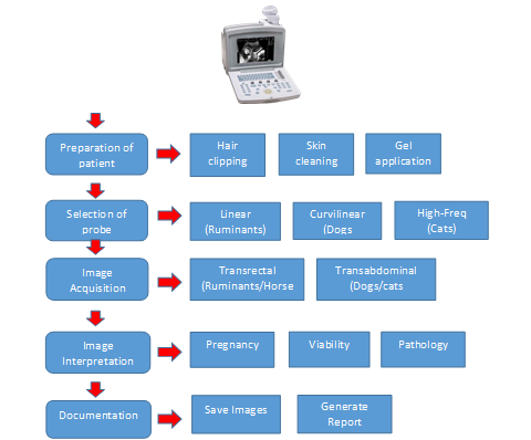
Figure 1. Flow Diagram of Veterinary Reproductive Ultrasonography
1.8. Evaluating the Safety of Diagnostic UltrasoundIn reproductive veterinary practice, ultrasound technology offers numerous diagnostic benefits for reproductive evaluation and monitoring of animals. Scientific evidence demonstrates that repeated ultrasound scanning procedures are biologically safe, with no observed negative impacts [59]. This technique is considered safe for animals, operators, and surrounding personnel, enabling its use in various settings without requiring special safety precautions. As a non-invasive procedure, ultrasound is well tolerated by animals, allowing for repeated assessments. This makes it particularly useful to monitor disease progression, evaluate treatment responses, and refine scanning techniques [29].
Ultrasound is a form of non-ionizing energy and is not associated with X-rays, which can harm tissues due to their ionizing effects on living cells. The pulsed ultrasound used in diagnostic imaging and Doppler devices for fetal monitoring operates at low intensity and does not generate significant heat. However, certain Doppler devices utilize higher intensities which can lead to considerable heating, making them unsuitable for fetal assessments. Another biological effect of ultrasound is cavitation, a complex phenomenon where gas-filled bubbles enlarge within the ultrasound field. At elevated intensities, these bubbles can suddenly collapse, causing localized temperature surges, thermal decomposition of water, and the generation of free radicals. This process is referred to as transient cavitation [64, 65]. Diagnostic ultrasound contrast agents have been created to improve echogenicity [66]. However, the biological effects of contrast-enhanced diagnostic ultrasound occur on a microscopic level and their clinical significance requires further research [64].
In comparison to other diagnostic tools like X-rays, ultrasound is widely regarded as very safe, with no harmful biological effects. However, the potential long-term biological impacts of ultrasound at diagnostic levels remain uncertain and necessitate further research.
2. CONCLUSION
To summarize, veterinary reproductive ultrasonography has emerged as a transformative tool, revolutionizing the diagnosis, monitoring, and management of reproductive health in animals. Recent advances in imaging technology, such as high-resolution probes and Doppler imaging, have significantly enhanced its precision and application. Future prospects lie in integrating artificial intelligence, portable devices, and advanced imaging modalities to further refine its capabilities. Despite remarkable advancements in veterinary reproductive ultrasonography, several research gaps persist, limiting its broader applicability and clinical precision across species and reproductive scenarios, such as doppler ultrasonography, 3D/4D ultrasound, and elastography are all well-established in human medicine but not widely adopted in veterinary reproduction. Furthermore, AI-assisted image analysis is still in early stages and there is a need for large, annotated veterinary ultrasound datasets to train AI models. Most studies focus on females, with fewer focusing on testicular pathologies, semen quality prediction, and breeding soundness exams using ultrasound, augmenting the need for standardized scrotal ultrasound protocols in different male species. Reproductive ultrasonography in endangered species is still experimental. This technique would become more useful by addressing these gaps. Continued research and collaboration among veterinarians, researchers, and industry stakeholders is essential to unlock the full potential of this technology, ensuring its widespread adoption and contribution to animal health and productivity.
AUTHOR CONTRIBUTION
Sajad Ali Laghari: writing-original Draft. Qudratullah Kalwar: Supervision. Muhammad Mohsen Rahimoon: writing review & editing. Fayaz Hussain: conceptualization Fazul U Rahman Soomro: conceptualization. Taj Muhammad: conceptualization. Abdul Saboor: conceptualization. Iqrar Uddin: conceptualization.
CONFLICT OF INTEREST
The authors of the manuscript have no financial or non-financial conflict of interest in the subject matter or materials discussed in this manuscript.
<DATA AVAILABILITY STATEMENT
Data supporting the findings of this study will be made available by the corresponding author upon request.
FUNDING DETAILS
No funding has been received for this research.
GENERATIVE AI DISCLOSURE STATEMENT
The authors did not used any type of generative artificial intelligence software for this research.
REFERENCES
- Meomartino L, Greco A, Di Giancamillo M, Brunetti A, Gnudi G. Imaging techniques in veterinary medicine. Part I: radiography and ultrasonography. Eur J Radiol Open. 2021;8:e100382. https://doi.org/ 10.1016/j.ejro.2021.100382
- Mantziaras G, Luvoni GC. Advanced ultrasound techniques in small animal reproduction imaging. Reprod Domest Anim. 2020;55:17–25. https://doi.org/ 10.1111/rda.13587
- Lanza GM. Ultrasound imaging: something old or something new? Invest Radiol. 2020;55(9):573–577. https://doi.org10.1097/RLI.0000000000000682
- Abdullah R, Asmad K, Khadijah WW, Diyana AN. Ultrasound scanning as a technique in pregnancy diagnosis of Saanen breed at UniSZA dairy goat farm. J Agrobiotechnol. 2018;9(1S):2–11.
- Ali Z, Sohail M, Ameen Y, Hamidullah AI, Malik M. Ultrasonography: a tool for management of reproductive disorders in dairy cows. Vet Sci Res Rev. 2023;9(1):18–24. https://doi.org/ 10.17582/journal.vsrr/2023/9.1.18.24
- Mali AB, Mehar RM, Kapane SH, Hadimani MR. Scanning the future: veterinary reproductive ultrasonography. Anim Reprod Update. 2022;2(1):82–89. https:// doi.org/10.48165/aru.2022.1201
- Ott TL, Tibary A, Waqas M, Geisert R, Giordano J. Pregnancy establishment and diagnosis in livestock. Annu Rev Anim Biosci. 2025;13(1):211–232. http://doi.org/10.1146/annurev-animal-021022-032214
- Khand FM, Kachiwal AB, Laghari ZA, et al. Early pregnancy diagnosis and fetometry by real time ultrasonography in teddy goat. J Anim Health Prod. 2021;9(3):342–348. https://doi.org/10. 17582/journal.jahp/2021/9.3.342.348
- Bucca S. Use of ultrasonography in fetal development and monitoring. In: Reef VB, Gentile L, Cuervo-Arango J, eds. Atlas of Equine Ultrasonography. Wiley Blackwell; 2022:383–406. https://doi.org/10.1002/9781119514671.ch19
- Mali AB, Amle MB, Markandeya NM, Kumawat BL. Comparative efficacy of trans-rectal and trans-abdominal ultrasonography for early diagnosis of pregnancy and embryonic ageing in goats. Indian J Small Rumin. 2019;25(2):171–175. https://doi.org/10.5958/0973-9718.2019.00046.1
- Ali A, Derar DR, Abdel-Razek ARK. Ultrasonography for the detection of pregnancy and study of embryonic and fetal development in camels, buffaloes, and sheep: techniques, equations, and limitations. Anim Reprod Sci. 2024;255:e107566. https://doi.org/ 10.1016/j.anireprosci.2024.107566
- Özdil A, Yılmaz O. Reproductive doppler ultrasonography practice in bitches. In: Kabu M, Tunç AC, eds. Current Research and Treatment in Animals. 2024;133–147. Livre De Lyon.
- Roos J, Aubanel C, Niewiadomska Z, Lannelongue L, Maenhoudt C, Fontbonne A. Triplex Doppler ultrasonography to describe the uterine arteries during diestrus and progesterone profile in pregnant and non-pregnant bitches of different sizes. Theriogenology. 2020;141:153–160. https://doi.org/10.1016/j.theriogenology.2019.08.035
- Simon S, Ramanathan A, Ghosh KN. Doppler ultrasonographic assessment of maternal and foetal blood flow in canine pregnancy and its application in the critical management of gestation. Indian J Anim Res. 2022;56(7):811–821. https://doi.org/10.18805/IJAR.B-4286
- Saha A, Mathur M. Ultrasound physics & overview. In: Li J, Chow RM-D, Vadivelu N, Kaye AD, eds. Ultrasound Fundamentals: An Evidence-Based Guide for Medical Practitioners. 2021:3–16. https://doi.org/10.1007/978-3-030-46839-2_1
- Bagley JE, Richter MP, Lane TJ. The role of transrectal sonography in pregnancy diagnosis in cattle. J Diagn Med Sonogr. 2023;39(1):50–60. https://doi.org/10.1177/87564793221120260
- Patey SJ, Corcoran JP. Physics of ultrasound. Anaesth Intensive Care Med. 2021;22(1):58–63. https://doi.org/10.1016/j.mpaic.2020.11.012
- Nielsen MB, Søgaard SB, Andersen SB, et al. Highlights of the development in ultrasound during the last 70 years: a historical review. Acta Radiol. 2021;62(11):1499–1514. https://doi.org/10.1177/02841851211050859
- Poulose BK. Ultrasonography basics. In: Docimo S, Blatnik JA, Pauli EM, eds. Fundamentals of Hernia Radiology. Cham, Switzerland. Springer International Publishing; 2023:21–27. https://doi.org/10. 1007/978-3-031-21336-6_3
- Gjesteby LA, Pare JR, Brattain LJ. Ultrasound for the emergency department and prehospital care. In: Cibis T, McGregor C, eds. Engineering and Medicine in Extreme Environments. Springer International Publishing; 2022:209–234. https://doi.org/10.1007/978-3-030-96921-9_11
- Duck FA, Baker AC, Starritt HC, eds. Ultrasound in Medicine. Boca Raton, FL: CRC Press; 2020.
- Stewart KA, Navarro SM, Kambala S, et al. Trends in ultrasound use in low and middle income countries: a systematic review. Int J MCH AIDS. 2020;9(1):103–113. https://doi.org/10.21106/ijma.294
- Grogan SP, Mount CA. Ultrasound Physics and Instrumentation. StatPearls Publishing; 2023. https://www.ncbi.nlm.nih.gov/books/NBK570593/
- Jaśkowski JM, Sobolewski J, Wieczorkiewicz M, Gehrke M, Herudzińska M. Modern techniques of teaching bovine rectal palpation: opportunities, benefits and disadvantages of new educational devices. J Vet Med Educ. 2020;47(1):5–10. https://doi.org /10.3138/jvme.2019-0012
- Balaro MFA, Cosentino IO, Ribeiro ACS, Brandão FZ. Ultrasound diagnosis in small ruminants: occurrence and description of genital pathologies. Vet Sci. 2022;9(11):e599. https://doi.org/10.3390/vetsci9110599
- Chandolia RK. Applications of transabdominal ultrasonography in bovine reproduction: a review. Buffalo Bull. 2022;41(2):225–240.
- Fulton RM. Focused ultrasound of the fetus, female and male reproductive tracts, pregnancy, and dystocia in dogs and cats. Vet Clin North Am Small Anim Pract. 2021;51(6):1249–1265. https://doi.org/10.1016/j.cvsm.2021.07.001
- Afzal S, Zahid M, Rehan ZA, et al. Preparation and evaluation of polymer-based ultrasound gel and its application in ultrasonography. Gels. 2022;8(1):e42. https://doi.org/10. 3390/gels8010042
- Mattoon JS, Sellon RK, Berry CR. Small Animal Diagnostic Ultrasound. 4th ed. St. Louis, MO: Elsevier; 2020.
- Szenci O. Recent possibilities for the diagnosis of early pregnancy and embryonic mortality in dairy cows. Animals. 2021;11(6):e1666. https://doi.org/10.3390/ani11061666
- Satheshkumar S. Application of ultrasonography in bovine reproduction. In: Kumaresan A, Srivastava AK, eds. Frontier Technologies in Bovine Reproduction. Singapore: Springer Nature Singapore; 2022:9–45. https://doi.org /10.1007/978-981-19-3072-0_2
- Alkan H, Kivrak MB, Satılmış F, Tekindal MA, Dinç DA. Detection of twin pregnancies in ewes by pregnancy-associated glycoprotein assay and transabdominal ultrasonography. Domest Anim Endocrinol. 2020;72:e106399. https://doi.org/10.1016/j.domaniend.2019.106399
- Christiansen D. Examination for pregnancy: Rectal palpation. In: Hopper RM, ed. Bovine Reproduction. 2nd ed. Hoboken, NJ: Wiley-Blackwell; 2021:471–478. https:// doi.org/10.1002/9781119602484.ch38
- Gardón JC, Acero BÁ, López SR. The use of ultrasonography in female bovine reproduction. In: Gardón JC, Ambrojo KS, eds. Assisted Reproductive Technologies in Animals. Vol 2. Cham, Switzerland: Springer; 2025:3–69. https://doi.org /10.1007/978-3-031-87198-6_1
- Adams GP, Singh J. Ovarian follicular and luteal dynamics in cattle. In: Hopper RM, ed. Bovine Reproduction. 2nd ed. Hoboken, NJ: Wiley-Blackwell; 2021:292-323. https:// doi.org/10.1002/9781119602484.ch25
- Bajaj NK. Reproduction and kidding in goats. In: Rana T, ed. Trends in Clinical Diseases, Production and Management of Goats. Springer Nature; 2024:151–162. https:// doi.org/10.1016/B978-0-443-23696-9.00019-5
- Barrón-Bravo O, Avilés-Ruiz R, Fraga-Escamilla E, Bautista-Martínez Y. Reproductive processes in cows and the ultrasonography use. Abanico Vet. 2023;13:e2023. https://doi.org/ 10.21929/abavet2023.18
- Magalhaes HB, Canisso IF, Dell-Aqua JA Jr. The temporal associations of B-mode and power-Doppler ultrasonography, and ovarian steroid changes of the periovulatory follicle and corpus luteum during luteogenesis and luteolysis in jennies. J Equine Vet Sci. 2023;122:e104224. https:// doi.org/10.1016/j.jevs.2023.104224
- Yáñez U, Murillo AV, Becerra JJ, Herradón PG, Peña AI, Quintela LA. Comparison between transrectal palpation, B-mode and Doppler ultrasonography to assess luteal function in Holstein cattle. Front Vet Sci. 2023;10:e1162589. https://doi.org /10.3389/fvets.2023.1162589
- Sumiyoshi T, Endo N, Tanaka T, Kamomae H. No adverse effect of confirmation of ovulation by rectal palpation and ultrasonography after artificial insemination on formation, development, and function of the corpus luteum and conception rate in cows. J Reprod Dev. 2022;68(6):377–382. https://doi.org/10.1262/jrd.2021-122
- Fricke PM. Scanning the future - ultrasonography as a reproductive management tool for dairy cattle. J Dairy Sci. 2002;85(8):1918–1926. https://doi.org/10.3168/jds.S0022-0302(02)74268-9
- Gnemmi G, Gardón JC, Maraboli C. Ultrasonography in bovine gynecology. In: Gardón JC, Satué K, eds. Biotechnologies Applied to Animal Reproduction: Current Trends and Practical Applications for Reproductive Management. Boca Raton, FL: Apple Academic Press; 2020:21–40.
- Detti L, Francillon L, Christiansen ME, et al. Early pregnancy ultrasound measurements and prediction of first trimester pregnancy loss: a logistic model. Sci Rep. 2020;10(1):e1545. https://doi.org/10.1038/s41598-020-58114-3
- Samsar MA, Lakoues HT, Yeddou MN, Aouane ND. Early Pregnancy Diagnosis Methods in Cow [doctoral dissertation]. Algiers, Algeria: Ecole Nationale Supérieure Vétérinaire (ENSV); 2024. http://depot.ensv. dz:8080/jspui/handle/123456789/2909
- Ortega-Ferrusola CGA, Da Silva-Álvarez E, Ortiz-Rodríguez JM, et al. Clinical application of Doppler ultrasound in the diagnosis of fertility problems in equines. Rev Bras Reprod Anim. 2021;45(4):459–469. https://doi.org/10.21451/1809-3000.RBRA2021.061
- Ortega-Ferrusola C, Gómez-Arrones V, Martín-Cano FE, et al. Advances in the ultrasound diagnosis in equine reproductive medicine: new approaches. Reprod Domest Anim. 2022;57:34–44. https://doi.org /10.1111/rda.14192
- Panzani D, Cuervo-Arango J, Fanelli D. Ultrasonographic pregnancy diagnosis in the mare. In: Gardón JC, Ambrojo KS, eds. Assisted Reproductive Technologies in Animals Volume 1: Current Trends for Reproductive Management. Cham, Switzerland: Springer Nature; 2024:29–47. https://doi.org/10.1007/ 978-3-031-73079-5_2
- Squires E. Current reproductive technologies impacting equine embryo production. J Equine Vet Sci. 2020;89:e102981. https://doi.org /10.1016/j.jevs.2020.102981
- Abdelnaby EA, Alhaider AK, El-Maaty AMA, Ragab RS, Seida AA, El-Badry DA. Ovarian and uterine arteries blood flow velocities waveform, hormones and nitric oxide in relation to ovulation in cows superstimulated with equine chorionic gonadotropin and luteolysis induction 10 and 17 days after ovulation. BMC Vet Res. 2023;19(1):e205. https://doi.org/10. 1186/s12917-023-03692-3
- Uçmak ZG, Kurban İ, Uçmak M. The vascularity of preovulatory follicle: the colour-Doppler assessment and its predictive value in the early pregnancy outcome in Arabian mares. Vet Hekim Der Derg. 2020;91(2):104–109. https://doi.org/10.33188/vetheder.672255
- Dascanio JJ. Assessment of late-term fetal well-being. In: Dascanio J, McCue P, eds. Equine Reproductive Procedures. Hoboken, NJ: Wiley-Blackwell; 2021:273–276. https:// doi.org/10.1002/9781119556015.ch73
- Gao Y, Hannan MA, Murata K, Rajabi-Toustani R, Nambo Y. Ultrasonographic examination of equine fetal growth parameters throughout gestation in pony for equine-assisted therapy. J Vet Med Sci. 2022;84(1):74–81. https://doi.org/ 10.1292/jvms.21-0301
- Murase H, Endo Y, Tsuchiya T, et al. Ultrasonographic evaluation of equine fetal growth throughout gestation in normal mares using a convex transducer. J Vet Med Sci. 2014;76(7):947–953. https://doi.org/ 10.1292/jvms.13-0259
- Gonzalez SW, Espy BMK, Stefanovski D, Turner RM. Real-time transrectal ultrasonographic measurement of the fetal eye (vitreous body) to predict parturition in bucking horse mares. J Equine Vet Sci. 2025;145:e105345. https://doi.org /10.1016/j.jevs.2025.105345
- Lubusky M, Studnickova M, Skrivanek A, Vomackova K, Prochazka M. Ultrasound evaluation of fetal gender at 12-14 weeks. Biomed Pap Med Fac Univ Palacky Olomouc Czech Repub. 2012;156(4):324–329. https://doi.org/10.5507/bp.2012.022
- Medan M, Watanabe G, Absy G, et al. Early pregnancy diagnosis by means of ultrasonography as a method of improving reproductive efficiency in goats. J Reprod Dev. 2004;50(4):391–397.
- Renaudin C, Wensley F, Morgan J, Cassano J, Spriet M. Transrectal ultrasonographic assessment of the fetal proximal phalanx: a new tool to assess fetal age and bone development in horses. Theriogenology. 2024;226:167–172. https://doi.org /10.1016/j.theriogenology.2024.06.010
- Colloton J. Ultrasound evaluation of the female reproductive tract. In: Hopper RM, ed. Bovine Reproduction. 2nd ed. Wiley-Blackwell; 2021:486–508. https://doi.org/10.1002/ 9781119602484.ch40
- Medan MS, Abd El-Aty AM. Advances in ultrasonography and its applications in domestic ruminants and other farm animals reproduction. J Adv Res. 2010;1(2):123–128.
- Kim D, Son M, Jung D, Heo S, Kim M, Yi J. Economic impacts of ultrasonographic fetal sex determination on Hanwoo cattle profitability and market dynamics. Vet Sci. 2025;12(3):e201. https://doi.org 10.3390/vetsci12030201
- Fontes PL, Oosthuizen N. Applied use of Doppler ultrasonography in bovine reproduction. Front Anim Sci. 2022;3:e912854. https://doi.org /10.3389/fanim.2022.912854
- Rehman HU, Kanwal M, Zardari AA, et al. Ultrasonographic estimation of fetal gestational age in the second and third trimesters using various biometric parameters. J Popul Ther Clin Pharmacol. 2024;31(5):2121–2126. https://doi.org/10.53555 /jptcp.v31i5.7474
- Francis YM, Karunakaran B. Ultrasonographic estimation of the gestational age using the fetal kidney length in the second and third trimesters of pregnancy among South Indian antenatal women: a cross-sectional study. Cureus. 2023;15(6):e40032. https://doi.org/10. 7759/cureus.41172
- Quarato CMI, Lacedonia D, Salvemini M, et al. A review on biological effects of ultrasounds: key messages for clinicians. Diagnostics. 2023;13(5):e855. https://doi.org/10. 3390/diagnostics13050855
- Zhou Y, Cui J, Deng CX. Dynamics of sonoporation correlated with acoustic cavitation activities. Biophys J. 2008;94(7):L51–L53. https://doi.org/ 10.1529/biophysj.107.125617
- Sinagra L, Orlandi R, Caspanello T, et al. Contrast-enhanced ultrasonography (CEUS) in imaging of the reproductive system in dogs: a literature review. Animals. 2023;13(10):e1615. https://doi.org/10.3390/ani13101615