
| Review | Open Access |
|---|
HPLC-Based Elucidation of Tannins from the Tissue and Callus Culture Extracts of Selected Medicinal Plants |

|
|---|
![]() Madieha Ambreen1*,
Madieha Ambreen1*,
![]() Safdar Ali Mirza2,
Safdar Ali Mirza2,
![]() and Zahida Bano3
and Zahida Bano3
1School of Biology, Minhaj University, Lahore, Pakistan
2Department of Botany, Government College University, Lahore, Pakistan
3University of Doha for Science and Technology, Qatar
Background. Optimized HPLC profiling is a powerful and effective analytical tool to standardize plant samples and authenticate plant materials. In this study, three selected medicinal plants namely: Achyranthes aspera, Ipomoea hederacea, and Ocimum basilicum were subjected to callus induction following seedling, leaf, and stem germination.
Methods. The induced callus was subsequently dried, finely ground, and extracted using methanol and water for HPLC analysis. A validated procedure was employed to identify and separate the tannin content in seedling leaf, stem, and callus culture extracts. HPLC fingerprinting was performed using a Shimadzu LC-20A system with a retention time of 2.9 minutes at 270 nm. The aim was to ensure quality and consistency in tannin analysis across different plant parts and callus culture samples.
Results. The highest callogenic response occurred in A. aspera leaf explants on MS medium with 2.0 mg/L 2,4-D and 4.0 mg/L NAA, producing green, granular callus. The lowest was in I. hederacea stem explants with 0.5 mg/L 2,4-D and BAP, yielding brown, granular callus. O. basilicum leaf callus extract showed the largest sample area (9365.56) and tannin content (2.66), with superior precision in tannin analysis for O. basilicum and A. aspera (7.81).
Conclusion. HPLC profiling proved to be an accurate, efficient, and precise method for evaluating tannin content in selected plant samples. It is a crucial method to standardize the quality of medicinal plant compounds.
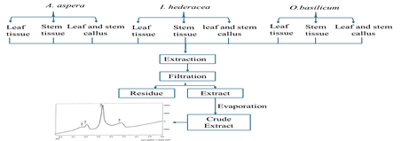
GRAPHICAL ABSTRACT
1. INTRODUCTION
Callus is a mass of mostly unorganized and undifferentiated cells. The plant tissue culture method provides a continuous and reliable source of natural products. The standardization of plant products is critical considering the growing demand for natural goods as medicines [1]. The production of secondary metabolites in cell culture relies on the amount/types of PGRs, carbon source, and other climatic conditions including light, temperature and gas composition [2].
Chromatographic fingerprinting presents a practical and effective solution to the global need for a stronger quality assessment method for traditional medicine [3]. This optimized technique enables the representation of chemical constituents distributed in plant materials, creating a “chemical database” that can be referenced in future research [4]. Tannins, also known as tannic acid, play an important biological role and have diverse applications. Their anticarcinogenic and antimutagenic effects are believed to stem from their antioxidant properties, which help protect cells from oxidative damage, including lipid peroxidation. Tannins also exhibit significant antibacterial activity. They suppress the growth of different microorganisms such as fungi, yeasts, bacteria, and viruses. Notably, propyl gallate and tannic acid but not gallic acid have been shown to inhibit the growth of aquatic bacteria, and microbes responsible for producing undesirable flavors [5].
The hydrolysis of ester linkages between gallic acid and polyols hydrolyzed during the maturing of any edible fruit is presumably linked to their antimicrobial capabilities. Tannins in these fruit, therefore, act as a natural barrier against microbial diseases [6]. Tannins demonstrate additional physiological effects, including lowering serum cholesterol, blood pressure, increasing blood coagulation and altering immune response [7]. The selected medicinal plants contain tannins, which have traditionally been used to treat a range of ailments. To demonstrate their effectiveness, it is essential to conduct both their qualitative analysis and quantification of tannin compounds present in various plant parts, using systematic scientific methods and comparisons with standard tannin compounds.
Achyranthes aspera L. belongs to the family Amaranthaceae. A. asperas is used to treat diarrhea, piles, heart disease, dyspepsia, vomiting, ascites, abdominal enlargement, and enlargement of the cervical gland [8-10]. Ipomoea hederacea of Convolvulaceae are known as ivy leaf morning glory or kaladana and habbunil. It is used to cure abdominal diseases, bronchitis, eye inflammation, gout, scabies, headache, constipation, fever, flatulence, leucoderma, eye disorders, splenopathy and hepatopathy [11]. Ocimum basilicum of Lamiacae/Labiatae is commonly known as basil or niazbo. It is used to treat stomachache and diarrhea due to its diuretic, demulcent and antipyretic properties [12]. While callus cultures provide a reliable source of secondary metabolites, there is limited standardized methodology employed for optimizing culture conditions with the aim to maximize tannin production. Moreover, despite the known medicinal potential of tannins in selected plants, comprehensive chromatographic profiling and quantitative analysis using HPLC remains underexplored for quality assurance and therapeutic validation. Hence, this study aims to use High Performance Liquid Chromatography (HPLC) to identify tannin compounds in selected medicinal plants by testing different mobile phase gradients and run times.
2. MATERIALS AND METHODS
2.1. Collection and Identification of Selected Plants
Healthy seeds of Achyranthes aspera L. (Voucher No. GC. Herb. Bot. 3492), Ipomoea hederacea (L.) Jacquin. (Voucher No. GC. Herb. Bot. 3493), and Ocimum basilicum L. (Voucher No. GC. Herb. Bot. 3491) were purchased from the market and identified by a taxonomist from GC University Lahore. The entire study was carried out at the Plant Biotechnology Laboratory, GC University of Lahore.
2.2. Seed Surface Sterilization and Callus InductionTo prevent microbial contamination during in vitro culture, seeds were subjected to sterilization using 3% sodium hypochlorite solution for 1 minute and then rinsed thrice with sterile distilled water. A total of 10 seeds from each species were aseptically placed in sterile Petri plates containing moist cotton pads to facilitate germination in the growth room.
Following successful germination, young seedling tissues (leaf and stem) were excised under sterile conditions and used for callus induction. Murashige and Skoog (MS) medium was supplemented with various concentrations of auxins (e.g.NAA, 2-4D) and cytokinins (BAP, KIN) to establish callus biomass production. Cultures were maintained at 25 ± 2°C under a 16-hour light/8-hour dark photoperiod provided by cool white; fluorescent lights to mimic optimal physiological conditions for callus development.
2.3. Drying and Maceration of CallusDeveloped callus tissues, along with the original leaf and stem tissues, were harvested and oven-dried at 40°C for one week to remove moisture while preserving bioactive compounds. Dried samples were ground into fine powder using a mechanical grinder. One gram of each powdered sample (leaf, stem, and corresponding callus) was weighed for tannin extraction, following the HPLC protocol outlined by reference [13].
2.4. HPLC profiling2.4.1. Preparation of Mobile Phase. A binary mobile phase consisting of methanol and water in a 1:1 ratio (50:50 mL) was prepared. The solvent mixture was thoroughly filtered using a 0.2 µm syringe filter to remove the particulate matter. Then, it was subjected to sonication for 20 minutes to ensure degassing and homogeneity, which improved peak resolution during chromatographic separation [14]
2.4.2. Preparation of Standard. Tannic acid was used as a standard and dissolved into 10 ml of mobile phase to form stock solution. A series of dilution (20 µg/ml, 30 µg/ml, 40 µg/ml and 50 µg/ml) was prepared and diluted with 10 ml of mobile phase. A calibration curve was plotted against the area.
2.4.3. Preparation of Sample. One gram of each plant tissue and macerated callus of leaf and stem were soaked in 10 mL of mobile phase and kept for 12 hrs with stirring. Then extracts were filtered using 0.2 µm syringe filters and subjected to 20 minutes of sonication to enhance solubility and remove trapped air. The resulting clear extracts were injected into the HPLC system for profiling and quantification.
Table 1. Chromatographic Instrumentation of Quantitative Determination of Tannins
|
Parameter |
Chromatograph Instrumentation |
|---|---|
|
HPLC system |
Shimadzu LC20A |
|
Injector |
Rheodyne |
|
Pump |
LC20AT |
|
Column |
A Hiber C18 Pore size 5µm. |
|
Mobile phase |
Methanol: water (50:50) |
|
Wavelength |
270 |
|
Flow rate |
20µl |
|
Rentation time |
2.9 min |
3. RESULTS
The leaf and stem explants of A. aspera, I. hederacea and O. basilicum were grown on MS media containing different combinations of plant growth regulators (PGRs). The effects of different concentrations of PGRs were recorded for the callogenic response of different explants of I. hederacea, that is, leaf, stem and root. The maximum callogenic response was exhibited by MS medium supplemented with 2.0 mg/l 2,4-D in combination with 4.0 mg/l NAA for the leaf of A. aspera as compared to other explants as the leaf callus had green colour and granular morphology (Figure 1A). The minimum callogenesis response was observed in MS medium supplemented with 0.5 mg/l 2,4-D, in combination with 0.5 mg/l BAP, for the stem of I. hederacea as compared to other explants. Stem callus was brown and had granular morphology. Stem explants of MSDB and MSDK of I. hederacea and O. basilicum respectively responded for callus induction with a low callus induction percentage (Table 2).
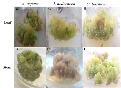
Figure 1. Effects of Different Concentrations of PGRs on Callus Induction (A) Leaf Explant of A. aspera on MSDN Medium (B) Stem Explant of A. aspera on MSDN Medium (C) Leaf Explant of I. hederacea on MSDB Medium (D) Stem Explant of I. hederacea on MSDB Medium (E) Leaf Explant of O. basilicum on MSDK Medium (F) Stem Explant of O. basilicum on MSDK Medium.
The combination of media is given in table 2 below.
Table 2. Effect of Different PGRs on Callus Induction of Different Parts of Selected Plants
|
Medium Code |
MS Media Compositions |
Explants |
Callus Induction (%) |
Callus Induction Duration (Days) |
Callus Index |
Callus Weight (g) |
Callus Morphology |
|---|---|---|---|---|---|---|---|
|
MSDN |
(2,4-D mg/l+ NAA mg/l ) 2.0+4.0 |
Leaf |
60 |
17 |
70 |
2.18±0.00 |
Green and granular |
|
(2,4-D mg/l+ NAA mg/l ) 6.0+8.0 |
Stem |
60 |
17 |
70 |
0.32±0.00 |
Green and compact |
|
|
MSDB |
(2,4-D mg/l + BAP mg/l) 0.5+1.5 |
Leaf |
40 |
17 |
200 |
0.56±0.03 |
Light green and granular |
|
(2,4-D mg/l + BAP mg/l) 0.5+0.5 |
Stem |
20 |
15 |
100 |
0.29±0.00 |
Brown and granular |
|
|
MSDK |
(2,4-D mg/l + KIN mg/l) 2.0+4.0 |
Leaf |
40 |
17 |
100 |
1.55±0.00 |
Light green and granular |
|
(2,4-D mg/l + KIN mg/l) 0.5+2.0 |
Stem |
20 |
14 |
60 |
0.39±0.005 |
Light green and granular |
3.1. HPLC-Based Comparison of Tannin Extraction from Leaf, Stem, and Callus Tissue of A. aspera, I. hederacea, and O. basilicum
Chromatogram of selected plants showed the retention time of 2.9 minutes at 270 wavelengths for tannin using methanol and water mobile phase. The tannins compound was analyzed for 5 minutes run time. The maximum area of sample was shown by O. basilicum callus culture extract, that is, 9365.56 among all explants and callus culture samples. The highest tannin content was exhibited by O. basilicum leaf callus culture extract, that is, 95.04 (Table 3).
Table 3. Quantitative Estimation of Tannin Content in Leaf and Stem of Selected Medicinal Plants
|
Plant Sample |
Appearance |
Area of Sample (a.u) |
Percentage of Tannins |
|---|---|---|---|
|
Achyranthes aspera leaf explants |
Brown |
2547.088 |
82.17 |
|
Achyranthes aspera leaf callus |
Dark brown |
3231.69 |
78.27 |
|
Ocimum basilicum leaf explants |
Brown |
7406.16 |
81.88 |
|
Ocimum basilicum leaf callus |
Dark brown |
9365.56 |
95.04 |
|
Iopomea hederacea leaf explants |
Light brown |
9287.89 |
83.16 |
|
Iopomea hederacea leaf callus |
Light brown |
18741.95 |
87.12 |
|
Achyranthes aspera stem explants |
Yellowish brown |
1241.350 |
93.06 |
|
Achyranthes aspera stem callus |
Dark brown |
8788.20 |
91.08 |
|
Iopomea hederacea stem explants |
Dark yellow brown |
3036.814 |
76.23 |
|
Iopomea hederacea stem callus |
Light brown |
4397.62 |
91.08 |
|
Ocimum basilicum stem explants |
Dark yellow |
6513.52 |
85.14 |
|
Ocimum basilicum stem callus |
Light yellow |
9291.56 |
81.18 |
aspera leaf seedling extract showed the highest area (1,245,086), indicating abundant bioactives. Ocimum basilicum leaf extract had the highest concentration (22.90 mg/mL), suggesting strong extractability. A. aspera stem tissue showed maximum recovery (135.45%), Further, its callus culture had the highest average signal (101.25), pointing to enhanced in vitro metabolite production. Conversely, Ipomoea hederacea stem seedling had the lowest area (41,976), indicating low phytochemical yield. The highest standard deviation (32.19) and RSD (23.97%) in I. hederacea extracts indicated low precision, requiring optimization (Table 4).
Table 4. HPLC Profiling of Selected Medicinal Plants
|
Extracts |
Area (mAU.s) |
Concentration recovered |
Recovery (%) |
Average |
STD |
RSD (%) |
|---|---|---|---|---|---|---|
|
A. aspera leaf seedling tissue extract |
1245086 |
74.30921219 |
99.07894959 |
85.85068182 |
11.81251798 |
5.282718638 |
|
A. aspera leaf callus (MSDN) extract |
8429952 |
20.31446254 |
125.4297502 |
101.5226771 |
14.47479398 |
6.814965447 |
|
I.hederacea leaf seedling tissue extract |
554041 |
11.91786102 |
95.34288814 |
49.19339767 |
30.93091877 |
13.8327274 |
|
I.hederacea leaf callus culture (MSDB) extract |
799874 |
18.93644721 |
75.74578884 |
37.65602505 |
32.18195984 |
14.39220997 |
|
O.basilicum leaf seedling tissue extract |
1386038 |
229.0315448 |
101.7917977 |
104.8058182 |
8.739828576 |
3.908570161 |
|
O.basilicum leaf callus culture (MSDK) extract |
365051 |
225.6780551 |
100.3013578 |
60.21710659 |
5.947601166 |
2.659848102 |
|
A. aspera stem seedling tissue extract |
668138 |
20.31446254 |
135.4297502 |
103.5226771 |
17.47479398 |
7.814965447 |
|
A. aspera stem callus culture (MSDN) extract |
563397 |
6.522154971 |
25.21772398 |
98.04397409 |
20.60680005 |
9.215641143 |
|
I.hederacea stem seedling tissue extract |
706150 |
16.70171649 |
111.3447766 |
99.42438963 |
53.55927427 |
23.95243562 |
|
I.hederacea stem callus culture (MSDB) extract |
439762 |
35.67155827 |
19.02483108 |
110.1427622 |
50.8640405 |
22.74709043 |
|
O.basilicum stem seedling tissue extract |
563397 |
24.87683255 |
82.92277516 |
95.54590879 |
15.47717828 |
6.921604547 |
|
O.basilicum callus stem callus culture (MSDK) extract |
497742 |
1386038 |
31.64734768 |
50.6357563 |
70.91843929 |
7.814965447 |
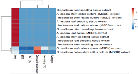
Figure 2. HPLC Profile of Standard Drug (Tannic Acid)
HPLC chromatograms at 270 nm display the tannin profiles extracted from the leaf, stem, and callus tissues of Achyranthes aspera, Ipomoea hederacea, and Ocimum basilicum, revealing distinct differences in the tannin content across tissue types and species. In all three plants, leaf extracts consistently exhibited the highest and sharpest peaks, particularly around the retention time of ~2.6 minutes, indicating a greater concentration of tannins (Figure 3). Stem extracts showed moderate peak intensities, reflecting lower tannin levels as compared to leaf tissues (Figure 4). In contrast, callus cultures displayed slightly reduced peak intensities, suggesting minimal tannin accumulation under in vitro conditions. Among the species, O. basilicum leaf extract showed the highest peak intensity, indicating it as the richest tannin source, followed by A. aspera and I. hederacea. These findings highlight that natural, differentiated tissues, especially leaves are superior to stem and callus tissues for tannin extraction and confirm species- and tissue-specific variation in tannin biosynthesis.
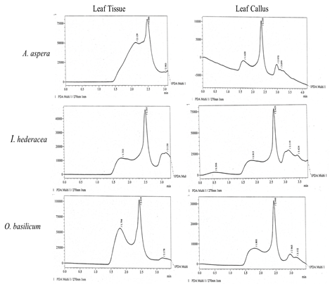
Figure 3. HPLC Profile of Seedling Leaf Tissue (Left Side) and Callus Culture (Right Side) of A. aspera, I. hederacea and O. basilicum. Leaf Extracts Exhibit Higher and Sharper Peaks, Especially Around ~2.6 Min, Indicating Greater Tannin Concentrations
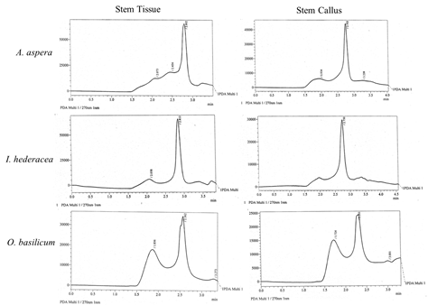
Figure 4. HPLC Profile of Seedling Stem Tissue (Left Side) and Callus Culture (Right Side) of A. aspera, I. hederacea and O. basilicum. Stem Extracts Show Moderate Peak Intensities
Figure 5. Comparative Analysis of the Heat Maps of HPLC Parameters of Selected Plants. The Colors Blue and Red Show the Value from the Lowest to the Highest, respectively.
The heat map illustrates the relative recovery or concentration of tannin compounds across various medicinal plant extracts, as analyzed by HPLC. Each row represents a specific extract from Ocimum basilicum, Achyranthes aspera, and Ipomoea hederacea, differentiated by plant part (leaf or stem) and tissue type (seedling tissue or callus culture). The color gradient from blue (low) to red (high) indicate the abundance of target compounds. Notably, the extracts from seedling tissues, particularly O. basilicum leaf and I. hederacea stem callus cultures, show higher compound recovery, while callus cultures, especially from A. aspera, generally exhibit lower recovery levels. This suggests that differentiated plant tissues may be more effective sources of bioactive tannins than callus cultures under the tested conditions (Figure 5).
4. DISCUSSION
The influence of different combinations of PGRs on the callogenic response of explants (leaf and stem) of the selected plants was assessed. Among the tested combinations, the maximum callus biomass production was noted in the leaf explants of Achyranthes aspera on MS medium fortified with 2.0 mg/L 2,4-D and 4.0 mg/L NAA. The resulting callus was green with a granular texture (Figure 1A), highlighting a robust morphogenic response as compared to other explants. Previous studies also highlighted the effectiveness of leaf explants in callus induction for A. aspera. Reference [15] reported successful callus formation using leaf explants cultured on MS medium supplemented with various concentrations of 2,4-D and NAA. The most pronounced callogenic response was achieved with a combination of 2 mg/L 2,4-D, IBA, BAP, IAA, and 4 mg/L NAA. Similarly, [16] also demonstrated efficient callus induction using leaf explants of A. aspera on MS medium enriched with varying concentrations of 2,4-D and NAA, further reinforcing the potential of this explant-PGR combination for the in vitro morphogenesis. In contrast, the lowest callogenic response was noted in stem explants of I. hederacea cultured on MS medium supplemented with 0.5 mg/L 2,4-D and 0.5 mg/L BAP. This callus exhibited a brown coloration with a granular morphology, indicating limited cellular proliferation. Callus initiation was typically observed within 13 to 17 days of incubation. Notably, stem explants from MSDB and MSDK variants of I. hederacea and O. basilicum demonstrated only modest callus induction, reflected by a relatively low callus formation percentage.
Chromatography, central to phytochemistry, is essential to obtain pure compounds for therapeutic development by enabling the separation, identification, and structural analysis of bioactive substances. It also plays a vital role in quality control and standardization of phytotherapeutics, primarily through HPTLC/HPLC fingerprinting and quantification of specific chemical markers. These techniques not only help in species identification and classification but also support the isolation and characterization of marker compounds, estimate genetic diversity, and offer real-time analyte detection through spectrum scanning and post-chromatographic derivatization[17-20].
HPLC settings were optimized to produce excellent chemical information and chromatograms with the best separation of neighboring peaks, which could be used to quantify the bioactive chemicals under investigation. The influence of different column temperatures, such as 25°C, 30°C, and 35°C on the separation process was also investigated. At 35°C, the majority of the peaks in HPLC chromatograms were well resolved. As a result, this temperature was chosen as the fingerprint analysis column temperature [21].
To examine the number of peaks and their spacing, detection wavelengths of 280, 254, and 360 nm were used. Finally, the wavelength of 280 nm was chosen, since this wavelength yielded more peaks than 254 or 360 nm. According to the current study, HPLC fingerprinting involves the characterization of bio active compounds. Mobile phase and column play an important role in isolating the compounds. Standards and extracts from seedling leaf, stem tissue, and callus cultures of selected plants showed a consistent baseline and a distinct peak characteristic of tannins, with 270 nm identified as the optimal wavelength for their detection. The tannin content of seedling leaf, stem tissue, callus culture extracts of selected plants exhibited a peak and retention time between 0 to 15 minutes. The HPLC profiling of seedling leaf, stem tissue and callus culture extracts of the leaf and stem of selected plants established a significant peak of tannin fraction at 2 to 2.9 retention time (Figure 3 and 4), (Table 1, 2). A previous study demonstrated that P. tuberosa showed best peak of tannic acid at 270 nm wavelength [22]. The presence of additional minor peaks further supports the occurrence of diverse polyphenolics, such as catechol and benzoic acid, as commonly reported in similar phytochemical analyses. Such findings were observed in the HPLC analysis of whole plant extracts of E. campestre, which revealed the presence of various polyphenolic compounds such as catechol and benzoic acid, further supporting the identification of tannins and related phenolics at the optimal detection wavelength of 270 nm [14]. The HPTLC analysis of ethanolic extracts from C. nilgirensis, C. gigantea, and C. crinita also revealed remarkable results, highlighting the presence of a diverse range of phytochemicals, including phenolics, flavonoids, and tannins. These results coincide with the current observations [23]. Another study [24] also confirmed the presence of polyphenols in the methanolic leaf extract of Synsepalum dulcificum. This finding also coincides with the current results.
aspera leaf tissue extract exhibited the highest chromatographic area (1,245,086), highlighting a rich abundance of bioactive compounds and confirming its potential as a valuable phytochemical source. In contrast, O. basilicum leaf extract recorded the highest concentration recovered (22.9031 mg/mL), suggesting it is chemically potent and highly extractable. Remarkably, A. aspera stem seedling tissue demonstrated the highest recovery percentage (135.45%), while its callus culture produced the strongest average signal (101.25), both pointing toward enhanced metabolite accumulation, likely due to favorable in vitro culture conditions stimulating biosynthesis. On the other hand, I. hederacea stem seedling tissue showed the lowest area (41,976), indicating a low phytochemical yield from that specific tissue. The highest standard deviation (32.19) and relative standard deviation (RSD) (23.97%) were observed for I. hederacea leaf callus and stem seedling extracts, respectively. These value reflected low analytical precision and signaled the need for optimization to improve reproducibility (Table 4).
The heat map shows that tannin compound recovery varied across different plant extracts, with higher levels found in seedling tissues especially in Ocimum basilicum leaves and Ipomoea hederacea stem callus cultures. In contrast, callus cultures, particularly from A. aspera, showed lower recovery. This indicates that differentiated tissues may be more suitable for extracting bioactive tannins, as compared to undifferentiated calluses under the conditions tested. These findings highlight the importance of tissue type in optimizing compound yield for medicinal plant research (Figure 5).
The HPLC fingerprints of these standard phenolic compounds acquired using the procedures outlined above might be used as standards for future research. Comparison with such standard chromatograms would provide both qualitative and quantitative analysis of the real phenolic compounds present in any unknown plant sample. This would allow the identification and confirmation of the presence of any of these 9 typical phenolic compounds in the study sample [25]. [26] used HPLC to extract condensed tannins from the bark of four tree species in Mexico; namely Arbutus xalapensis Kunth, Prunus serotina Ehrh., Quercus crassifolia Humb. & Bonpl, and Quercus laurina Humb. & Bonpl. In that study, the total tannin content was also isolated from the Citrus limon extract using HPLC, which aligns with the methodology employed in the current study.
4.1. ConclusionThe current HPLC data revealed distinct trends among species and tissue types. A. aspera emerged as a metabolically rich plant, especially in its seedling and stem tissues, while O. basilicum displayed exceptional reproducibility and high chemical recovery, particularly in its callus forms. I. hederacea, although showing potential, demonstrates higher variability, especially in its stem extracts. This profiling not only underscores the phytochemical potential of these plants but also identifies which culture conditions and plant parts yield the best results, paving the way for future standardized extraction and pharmacological applications.
Author Contribution
Madieha Ambreen: conceptualization, investigation, methodology, writing – original draft. Safdar Ali Mirza: supervision, visualization, writing – review & editing. Zahida Bano: software formal analysis, data curation.
Conflict of Interest
The authors declare that they have no conflict of interest regarding the publication of this manuscript.
Data Availability Statement
Data sharing is not applicable to this article as no new data were generated during the study.
Funding Details
No funding has been received for this research.
Generative AI Disclosure Statement
The authors did not used any type of generative artificial intelligence software for this research.
Acknowledgment
The authors express their gratitude to Dr. Zaheer-ud-din Khan from the Department of Botany, GC University Lahore, Pakistan, for his assistance in the identification of plant specimens
REFERENCES
- Steward FC, Mapes MO, Mears K. Growth and organized development of cultured cells. II. Organization in cultures grown from freely suspended cells. Am J Bot. 1958;45(9):705–708. https://doi.org/10.1002/j.1537-2197.1958.tb10599.x
- Sen MK, Nasrin S, Rahman S, Jamal AHM. In vitro callus induction and plantlet regeneration of Achyranthes aspera L., a high value medicinal plant. Asian Pac J Trop Biomed. 2014;4(1):40–46. https://doi.org /10.1016/S2221-1691(14)60206-9
- Haliński LP, Szafranek J, Szafranek BM, Gołębiowski M, Stepnowski P. Chromatographic fractionation and analysis of the main components of eggplant (Solanum melongena L.) leaf cuticular waxes. Acta Chromatograph. 2009;21(1):127–137. https://doi.org /10.1556/AChrom.21.2009.1.11
- Tiwari S, Bhadoriya U, Saini L, Gupta A, Solanki S. Quantitative analysis of glycyrrhizic acid by HPTLC in herbal formulation. Asian J Pharmaceut Life Sci. 2011;1(2):124–127.
- Winiarska-Mieczan A, Muszyński S, Tomaszewska E, et al. The impact of tannic acid consumption on bone mineralization. Metabolites. 2023;13(10):e1072. https://doi.org /10.3390/metabo13101072
- Choubey S, Goyal S, Varughese L, Kumar V. Probing gallic acid for its broad-spectrum applications. Mini Rev Med Chem. 2018;18(15):1283–1293. https://doi.org/10.2174/1389557518666180330114010
- Chung KT, Wong TY, Wei CI, Huang YW, Lin Y. Tannins and human health: a review. Critical Rev Food Sci Nutr. 1998;38(6):421–464. https:// doi.org/10.1080/10408699891274273
- Banerjee KGJ, Gupta AK, Daha P. Phytochemical constituents and pharmacological uses of medicinal plant Achyranthes aspera: a review. World J Pharmaceut Res. 2015;4(1):470–489.
- Shashikanth J, Dorcas M, Mugendhiran S, Renu. Biodiversity with special reference to indigenous systems of medicinal plants, ornamental and weeds of University College for Women, Koti, Osmania University, Hyderabad, Telangana, India. Ann Plant Sci. 2023;12(10):5976–6032. https://doi. org/10.21746/aps.2023.12.10.1
- Ambreen M, Mirza SA, Bano Z, et al. Antioxidant and anticancer activity of tannins isolated from callus cultures of Achyranthes aspera L. Sci Inq Rev. 2024;8(2):1–21. https://doi.org /10.32350/sir.82.01
- Haq MZU, Riaz M, De Feo V. Ipomea hederacea Jacq.: a medicinal herb with promising health benefits. Molecules. 2012;17(11):13132–13145. https:// doi.org/10.3390/molecules171113132
- Awan ZI, Habib-ur-Rehman A, Awan A, Minhas FA, Khan MN. Ethnobotanical importance of some highly medicinal plants of District Muzaffarabad, Pakistan with special reference to the species of the genus Viburnum. J Pharm Biol Sci. 2013;6(2):53–66. https://doi.org/ 10.9790/3008-0625366
- Tanveer H, Safdar A, Asi MR. Appraisal of an important flavonoid, quercetin, in callus cultures of Citrullus colocynthis. Int J Agric Biol. 2012;14(4):1814–9596.
- Al-Askar AAI, Bashir S, Abdallah E, et al. Antimicrobial efficacy and HPLC analysis of polyphenolic compounds in a whole-plant extract of Eryngium campestre. Separations. 2023;10(6):e362. https://doi.org/ 10.3390/separations10060362
- Senthilmanickam J, Bhavani AL, Venkatramlingam K, Chandra G. The role of 2,4-D and NAA in callus induction of Achyranthes aspera and its secondary metabolite studies. J Appl Nat Sci Online. 2012;2(3):232–243.
- Kayani S, Zia M, Sarwar S, Chaudhary MF. Callogenic studies of Achyranthes aspera leaf explant at different hormonal combinations. Pak J Biol Sci. 2008;11(6):950–952. https://doi. org/10.3923/pjbs.2008.950.952
- World Health Organization. The Selection of Essential Drugs. Second Report of the WHO Expert Committee. World Health Organization; 1979.
- Edeoga HO, Okwu DE, Mbaebie BO. Phytochemical constituents of some Nigerian medicinal plants. Afr J Biotechnol. 2005;4(7):685–688. https://doi.org/10.5897/AJB2005.000-3127
- Kpoviéssi DSS, Gbaguidi F, Gbénou J, et al. Validation of a method for the determination of sterols and triterpenes in the aerial part of Justicia anselliana (Nees) T. Anders by capillary gas chromatography. J Pharmaceut Biomed Anal. 2008;48(4):1127–1135. https://doi.org/10.1016/j.jpba.2008.08.036
- Faiyazuddin MDA, Rauf N, Ahmad. A validated HPTLC method for determination of terbutaline sulfate in biological samples: application to pharmacokinetic study. Saudi Pharmaceut J. 2011;19(3):185–191. https://doi.org/10.1016/j.jsps.2011.03.004
- Ahmed R. High-performance liquid chromatography (HPLC): principles, applications, versatility, efficiency, innovation and comparative analysis in modern analytical chemistry and in pharmaceutical sciences. Clinic Invest. 2024;14(9):524–535. https://doi.org/ 10.20944/preprints202409.0057.v1
- Durgawale TP, Durgawale PP, Khanwelkar CC. Quantitative estimation of tannin by HPLC. Der Pharm Lett. 2016;8(3):123–126.
- Mradu GS, Saumyakanti, Sohini M, Arup M. HPLC profiles of standard phenolic compounds present in medicinal plants. Int J Pharm Phytochem Res. 2012;4(3):162–167.
- Obafemi TO, Akinmoladun AC, Olaleye MT, et al. High performance liquid chromatography (HPLC) fingerprinting, mineral composition and in vitro antioxidant activity of methanol leaf extract of Synsepalum dulcificum (Sapotaceae). J Appl Pharmaceut Sci. 2017;7(10):125–131.
- Ruiz-Aquino F, Feria-Reyes R, Rutiaga-Quiñones JG, Robledo-Taboada LH, Gabriel-Parra R. Characterization of tannin extracts derived from the bark of four tree species by HPLC and FTIR. Forest Sci Technol. 2023;19(1):38–46. https://doi.org/10.1080/21580103.2023.2166593
- Rathod ZR, Sarita S, Saraf MS. Identification and estimation of total tannins from Citrus limon L. Burm. f. (lemon) and its endophytes. Curr Trends Biomed Eng Biosci. 2022;20(5):1–12.
Steward et al. demonstrated that freely suspended plant cells in culture can develop organized structures, highlighting the totipotency of plant cells. This study laid foundational work for modern plant tissue culture and regeneration techniques.
Sen et al. optimized in vitro protocols for callus induction and plantlet regeneration of Achyranthes aspera, a medicinally important plant. Their findings support conservation and large-scale propagation through tissue culture techniques.
Haliński et al. analyzed the chemical composition of cuticular waxes from Solanum melongena leaves using chromatographic techniques. Their study identified major wax components, contributing to the understanding of plant surface chemistry and defense.
Tiwari et al. performed quantitative analysis of glycyrrhizic acid in herbal formulations using High-Performance Thin Layer Chromatography (HPTLC). The study ensured quality control and standardization of herbal products containing Glycyrrhiza glabra.
Winiarska-Mieczan et al. investigated the effects of tannic acid consumption on bone mineralization. Their findings revealed that excessive intake may negatively influence bone health, highlighting potential dietary risks.
Choubey et al. reviewed the diverse pharmacological applications of gallic acid, emphasizing its antioxidant, antimicrobial, and anticancer properties. The study highlights its potential as a multifunctional bioactive compound in medicinal chemistry.
Chung et al. provided a comprehensive review on tannins, discussing their sources, biological activities, and effects on human health. The study highlighted both beneficial and adverse impacts, depending on dosage and dietary context.
Banerjee et al. reviewed the phytochemical profile and pharmacological activities of Achyranthes aspera, emphasizing its therapeutic potential. The study compiles traditional and modern uses, supporting its role in herbal medicine.
Shashikanth et al. documented the biodiversity of medicinal, ornamental, and weed plant species at University College for Women, Osmania University. The study emphasizes the importance of indigenous plant systems for conservation and sustainable use.
Ambreen et al. evaluated tannins extracted from Achyranthes aspera callus cultures for their antioxidant and anticancer properties. The study demonstrated significant bioactivity, supporting their potential use in therapeutic applications.
Haq et al. reviewed the medicinal properties of Ipomoea hederacea, highlighting its phytochemical composition and potential therapeutic benefits. The study supports its traditional use and encourages further pharmacological research.
Awan et al. explored the ethnobotanical significance of medicinal plants in Muzaffarabad, Pakistan. The study highlights traditional knowledge and the therapeutic potential of these plants for local healthcare.
Tanveer et al. assessed the presence of quercetin, a valuable flavonoid, in callus cultures of Citrullus colocynthis. The study underscores the potential of in vitro cultures for producing bioactive compounds with medicinal value.
Al-Askar et al. investigated the antimicrobial activity and polyphenolic profile of whole-plant extracts of Eryngium campestre using HPLC. The study revealed strong antimicrobial efficacy, highlighting its potential in natural therapeutics.
Senthilmanickam et al. examined the effects of 2,4-D and NAA on callus induction in Achyranthes aspera and analyzed its secondary metabolites. The study demonstrated hormone-specific responses influencing callus formation and bioactive compound production.
Kayani et al. investigated callus induction in Achyranthes aspera leaf explants using various hormonal combinations. The study identified optimal conditions for callogenesis, aiding in tissue culture-based propagation of the species.
WHO published the second report on the selection of essential drugs, outlining criteria for prioritizing medicines based on public health relevance, efficacy, and cost-effectiveness. This report laid the groundwork for national essential medicines lists worldwide.
Edeoga et al. analyzed the phytochemical constituents of selected Nigerian medicinal plants, identifying key compounds like alkaloids, flavonoids, and tannins. The study supports the therapeutic potential of these plants in traditional medicine.
Kpoviéssi et al. validated a capillary gas chromatography method for analyzing sterols and triterpenes in the aerial parts of Justicia anselliana. The study ensures accurate phytochemical profiling for quality control in herbal formulations.
Faiyazuddin et al. developed and validated an HPTLC method for detecting terbutaline sulfate in biological samples. The method was successfully applied in pharmacokinetic studies, offering a reliable tool for drug monitoring.
Ahmed provided an in-depth overview of High-Performance Liquid Chromatography (HPLC), highlighting its principles, efficiency, and wide-ranging applications in analytical chemistry and pharmaceutical sciences. The study emphasizes HPLC's innovation and versatility in modern research.
Durgawale et al. conducted quantitative estimation of tannins using HPLC, demonstrating an accurate and efficient method for tannin analysis. The study supports its application in standardizing tannin-rich herbal formulations.
Mradu et al. presented HPLC profiles of standard phenolic compounds in various medicinal plants, enabling accurate identification and quantification. The study aids in quality control and validation of plant-based formulations.
Obafemi et al. performed HPLC fingerprinting and mineral analysis of Synsepalum dulcificum leaf extract, revealing significant antioxidant activity. The study supports its pharmacological potential and use in herbal medicine.
Ruiz-Aquino et al. characterized tannin extracts from the bark of four tree species using HPLC and FTIR techniques. Their findings provided detailed insights into the chemical composition and potential applications of these natural extracts.
Rathod et al. identified and quantified total tannins from Citrus limon and its associated endophytes. The study highlights the role of endophytes in enhancing bioactive compound production for potential therapeutic applications.