
| Review | Open Access |
|---|
Therapeutic Potential of Graphene Nanoparticles in Breast Cancer: Insights from the Past Ten Years |
 |
|---|
![]() Ishrat Younus1*, Afshan Siddiq2, Sarah Jameel Khan1, Nimra Mujeeb1, and Rafia Sadaf3
Ishrat Younus1*, Afshan Siddiq2, Sarah Jameel Khan1, Nimra Mujeeb1, and Rafia Sadaf3
1Faculty of Pharmacy, Hamdard University, Karachi, Pakistan
2Faculty of Pharmacy and Pharmaceutical Sciences, University of Karachi, Karachi, Pakistan.
3Faculty of Pharmacy, Jinnah College of Pharmacy, Sohail University, Karachi, Pakistan
Breast cancer is the second leading cause of death in women all over the world and its prevention is still a challenge. There are various systemic and local therapies used in the treatment of breast cancer. These therapies, including chemotherapy, endocrine treatment, and tissue-targeted therapies, complement definitive local treatments including surgery or radiation, leading to a significant reduction in both cancer recurrence and disease-related mortality. However, traditional cancer therapies are limited in their specificity and systemic toxicity. In this scenario, nanomaterials may prove to be valuable in providing effective cancer treatments, while reducing unwanted side effects and providing precise diagnosis. Graphene nanoparticles and its derivative reduced graphene oxides (RGO), which belong to a category of carbon nanomaterial, have captured significant interest for their matchless utility as fundamental components in breast cancer treatment. This review focuses on different types of graphene-based nanomaterials which exhibit significant potential in drug delivery applications due to their unique properties, including high drug-loading capacity, pH-dependent release, and enhanced cellular uptake. A thorough search of a bibliographic database during the last decade (2013-2023), was conducted to find the research articles focused on nanotechnology using graphene for cancer treatments to gather the relevant information.
Highlights
- NE-based formulations of AZM were developed using essential oils to enhance its solubility and stability.
- Peppermint oil and Tween 20 demonstrated the highest emulsification efficiency and transmittance.
- Comprehensive characterization confirmed stability through thermodynamic stability, viscosity, refractive index, and particle size analysis.
- The potential for improved drug delivery with enhanced solubility and bioavailability of AZM is highlighted.
1. INTRODUCTION
Nanotechnology has continuously improved the survival rate of cancer patients as well as the treatment protocols used to stop the recurrence and proliferation of cancerous cells. The cytotoxic effects on normal cellular growth and efficient delivery of drugs for targeted treatment have been incorporated into nanoparticles based system. In achieving the desirable goals, such as improving survival rates of cancer patients, preventing recurrence and proliferation of cancer cells, minimizing cytotoxic effects on normal cells and enhancing efficiency of targeted drug delivery, the role of carbon nanoparticles is noteworthy, especially of a novel graphene-based nanoentity, because of its physicochemical properties. Graphene is being used in anti-cancer therapeutic regime with its targeted delivery, its nanoconjugates, its pH-controlled releasing abilities, and for its contribution in the early diagnosis of breast cancer. This nanoparticle can be further engineered to reduce its side effects and enhance treatment efficacy [1]. Graphene can be helpful for the diagnosis of breast cancer cells in their initial stages.
For the preliminary identification of breast cancer, an electrochemical procedure is performed by using gold nanomaterial and graphene oxide over glassy carbon electrodes. The gold nanomaterial system enables enhanced electrical and chemical signal generation, leading to a greater electrochemical charge difference. Even the lowest detection limit is achieved with high reproducibility [2].
Graphene nanoparticle helps in the identification of the biomarkers for breast cancer. For this purpose, many techniques are utilized along with the use of graphene oxide, such as Raman spectroscopy, Fourier transform infrared spectroscopy, scanning electron microscopy, UV spectroscopy, and x-ray spectroscopy [3].
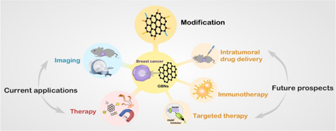
Figure 1. Graphene Nanoparticles: Current Applications and Future Prospects
Previous studies showed that graphene oxide nanosystems have the ability to kill tumor cells and can be loaded with a large number of drugs due to their high surface area and surface functionality. These properties make graphene nanosystems effective for use against breast cancer [4]. Similarly, a study done by Masoumzade highlighted some properties of graphene quantum dots (water solubility, low toxic effects, chemically inert, high biocompatibility, fluorescence ability, and increases the penetration ability of other cancer drugs), due to which they are used in the control and treatment of breast cancer [5]. Another study highlighted similar aspects of graphene oxide nanosystems, such as their high surface area, low toxicity, and distinctive physicochemical and mechanical properties, which make them powerful anti-cancer agents [6].
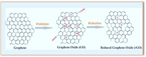
Figure 2. Conversion of Graphene Nanoparticles to Reduced Graphene Oxide
This review provides a comprehensive and focused analysis of the therapeutic and diagnostic potential of graphene nanoparticles, specifically targeting breast cancer. Unlike previous reviews, it categorizes various graphene-based systems, such as pure graphene, graphene oxide (GO), reduced graphene oxide (rGO), graphene quantum dots (GQDs), and their functionalized forms with drugs, herbs, chemicals, and metals. This approach allows for a detailed comparison of recent advancements in drug delivery systems, photothermal therapy, biosensing, and their synergistic application when integrated with conventional treatments. Additionally, this review discusses emerging trends and recent studies published in 2023, addressing challenges and proposing future perspectives. Given the rapid advancements in the field, recent studies have been critically discussed to reflect the most up-to-date trends aimed at enhancing the clinical efficacy of graphene-based nanomaterials against breast cancer.
2. GRAPHENE WITH DRUGS
2.1. Graphene with MitoxantroneSome gold nanoparticles are combined and modified with graphene oxide to create a covalent drug delivery system aimed to efficiently transport anti-cancer drug molecules to breast cancer cells. Mitoxantrone is an anti-cancer medicine, modified with gold particles and mercaptopropionic acid spacer (SMTX-AuNPs). In this study, this system was compared with mercaptopropionic acid with gold particles and nanoparticles combined with graphene oxide (MPS-AuNPs/GO) by using a similar technique [2]. Previous research suggested that the combination of a chemotherapeutic agent (mitoxantrone), transforming growth factor beta (TFG-β), and reduced graphene oxide (rGO) stopped distant metastasis, damaged local cancer cells, and improved the immunosuppressive microenvironments in mice, when administered through in situ vaccine [7]. In vitro studies were performed to observe the efficacy of drug delivery. The results indicated that engineered nanoparticles were more efficient as compared to free drugs [2].
2.2. Graphene with DoxorubicinThe chemotherapeutic results indicated that the activity of breast cancer cells was reduced by up to 50%, which is significantly greater than that observed with free and uncoated doxorubicin. In addition, nanosystem didn’t show any signs of cytotoxicity to normal cellular growth. Thus, this nanocomposite is functional and useful for the targeted delivery of drugs in cancerous cells [7].
Astani et al. [8] developed doxorubicin and cisplatin loaded reduced graphene nanocomposite, which exhibited higher cytotoxic effects even at a low concentration in MCF-7 breast cancer cell line. In one study, researchers found significantly greater cytotoxic effect of graphene oxide and doxorubicin on two human breast cancer cell lines (MCF-7 and BT474) in comparison to the application of doxorubicin and graphene oxide alone [9].
2.3. Carboxylate Graphene Oxide (cGO) with Doxorubicin and SilibininThe combined effect of DOX and silibinin was examined against two breast cell lines, namely MCF-7 and SK-BR-3. A drug delivery carrier was successfully developed by coating carboxylated graphene oxide (cGO) with a HB5 aptamer, lowering the CDK2, mTOR, Akt, and NF-B levels by increasing Rb levels and accelerating the apoptosis of cells. The created Apt-cGO-DOX-Sili can be recommended as an easy and effective drug delivery method for breast chemotherapy [10].
2.4. Functionalized Graphene Nanosheets for DOX DeliveryNanotechnology has emerged as a promising approach to overcome the challenges associated with chemotherapy. A specialized carbon nanosheet was designed to treat breast cancer cells by delivering doxorubicin. These nanosheets were coated with multi-cyclodextrin. Various tests, such as XRD, MAP, EDS, FTIR, VSM, SEM, and TEM were used to assess the structural features of the nanocarrier. MTT and hemolysis assays were used to monitor the biocompatibility of the synthesized drug-carrier against MCF-7 cells. These results showed the potential of doxorubicin to treat breast cancer cells [11].
2.5. Graphene Oxide with Doxorubicin and GinsenosideThe conjugates of GO-Rg3-DOX were used to inhibit the growth of MDA-MB-231 breast cancer cells and Huh7 hepatocarcinoma. Due primarily to the activation of apoptotic genes and the down regulation of transcription regulatory genes, GO-Rg3-DOX conjugates drastically decreased the cancer cell viability. GO-Rg3 is a DOX carrier that is efficient, biocompatible, pH-responsive, and has the potential to enhance chemotherapy, at least when used to treat breast and liver malignancies [12].
2.6. Graphene Oxide Nanoplatform to Boost Cisplatin-Based Drug Delivery in Anti-cancer TherapyPlatinum-based drugs are commonly used to treat various cancers; however, their application is limited due to their several side effects, including a high rate of drug degradation before cellular uptake, the development of drug resistance, and off-target organ toxicity. These limitations in drug delivery can be overcome by designing a nanocarrier that could be used to increase the accumulation of drugs in targeted cells in order to decrease drug toxicity. Researchers have synthesized a highly versatile 2D nanoplatform composed of graphene oxide for the use of lower amounts of platinum drugs, as compared to a platinum-free complex. This nanoplatform possesses a high inhibition potential for cellular proliferation and migration in osteosarcoma [13].
2.7. Graphene Oxide with Doxorubicin and MethotrexateIn a previous study, a dual drug-loaded transdermal drug therapy was used to treat the cancer, consisting of anionic graphene oxide, cationic polyethyleneimine, and polyanionic dextran sulphate (GO/PEI/DS). The results showed that this synthesized dual drug-loaded material has a good pH-dependent profile for the transdermal delivery of both MTX and DOX in comparison with oral delivery [14].
3. GRAPHENE WITH CHEMICALS
3.1. Graphene with Chondroitin Sulfate Multialdehyde or PolyethylamineIt also helped to reduce breast cancer reoccurrence in some cases after surgery. The procedure included chondroitin sulfate multialdehyde, polyethylamine, and its combination with graphene particles. The stabilized structure facilitated the targeted delivery of the drug in vitro into breast cancer cells. Additionally, the prevention of tumor recurrence was observed in murine models [15].
3.2. Graphene with Cobalt Tetra Phenoxy Acetic Acid PhthalocyanineIn this study, cobalt tetra (phenoxy acetic acid) phthalocyanine (CoTAPc) was utilized to enhance the electrochemical signal for the detection of human epidermal growth factor receptor 2 (HER2). The aptasensors demonstrated optimal sensitivity, specificity, and storage stability for HER2 detection, highlighting their strong potential for early-stage breast cancer diagnosis [16].
3.3. Graphene with Cationic PolymerIn this research, the cationic polymer poly(methacrylic acid-co-diallyl dimethyl ammonium chloride) (PMA-DDA), coated on GQDs, was molecularly imprinted with the chemotherapy drug doxorubicin (DOX). Fourier-transform infrared spectroscopy (FT-IR) was used to examine and confirm the chemical structure of PMA DDA-coated GQDs. The images revealed that PMA DDA-coated GQD particles had gathered around the tumor cells [12].
3.4. Graphene Oxide with Polyamidoamine DendrimerThis study aimed to explore the possibility of a graphene oxide (GO) based nanocarrier coated with poly (amido amine) dendrimers for the targeted delivery of quercetin (QSR), a hydrophobic anti-cancer medication. The study emphasizes the potential use of synthetic hybrid materials as a nanocarrier for the delivery of a hydrophobic anti-cancer medication with great loading and controlled release efficiency [17].
4. GRAPHENE WITH METALS
4.1. Graphene with Manganese-Zinc SulfideThe role of nanoparticles in cancer therapy is significant for targeted drug delivery and treatment but their dispersibility remains poor. To overcome this issue, graphene oxide in its reduced form was coupled with manganese-zinc sulfide and activated with folic acid. The comparison was based on doxorubicin activity, its specificity, non-toxic nature, and compatibility with the physiological system. The chemotherapeutic conclusion presented that the activity of breast cancer cells was diminished up to 50%, which is significantly greater than with free and uncoated doxorubicin. In addition, the nanosystem didn’t show any signs of cytotoxicity to normal cellular growth. Moreover, the nanocomposite was found to be functional and useful for the targeted delivery of drugs in cancerous cells [7].
4.2. Graphene with PlatinumTo prevent chemoresistance, graphene nanoparticles were combined with platinum and their functions were analyzed with respect to various genetically mutated genes and other cancerous cell cultures. Their drug delivery to cancerous cells was effectively observed through assays, morphological changes, and via the examination of apoptotic markers. The activity of graphene-based conjugate with platinum was investigated and found to be more effective as therapy. This nanocomplex exceptionally enhanced the transportation and distribution of anti-cancer drugs [18].
4.3. Graphene Oxide with Titanium Carbide MXene (Ti3C2)The radiosensitivity of breast cancer cells was examined utilizing clinical x-ray irradiation to determine the effectiveness of two carbon-based nanomaterials, that is, graphene oxide and Ti3C2 MXene. The results demonstrated that Ti₃C₂ MXene nanoparticles act as more effective radiosensitizers compared to graphene oxide [19].
4.4. Graphene Oxide with Cerium OxideIn the current study, a cerium oxide/graphene oxide hybrid (CeO2/GO hybrid) nanocomposite system was created using the ultrasonic method. The MCF-7 cell line’s CeO2/GO hybrid revealed an increase in the apoptotic response as sample concentration increased. Hence, the CeO2/GO platform can be employed as a treatment platform for breast cancer. The generated CeO2/GO hybrid’s synergistic effects with the MCF-7 cell line are shown below in Figure 3 [20].
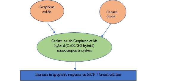
Figure 3. Synergistic Effects of Graphene Oxide and Cerium Oxide on Breast Cell Lines
4.5. Graphene with Tin SulfideIn the current study, an enzyme-free sensor was designed and built utilizing N-doped graphene quantum dots (N-GQDs)-decorated tin sulfide nanosheets (SnS2). Lastly, the N-GQDs@SnS2 electrode was successfully utilized to monitor the H2O2 produced in real-time from mouse fibroblasts and breast cancer cells. This opens the door for the development of efficient non-enzymatic electrochemical sensors for the detection of different biomolecules applying a simple technique [21].
4.6. Graphene Oxide with Copper and Nickel Spinel FerriteThis study evaluated how differently graphene oxide (GO), chemically reduced graphene oxide (rGO), and copper and nickel spinel ferrite nanoparticles (CuFe2O4 NPs and NiFe2O4 NPs) behave. In comparison to NiFe2O4 NPs, cytotoxicity was reduced in HepG2 and T98G cell lines treated with rGO-NiFe2O4 NCs, while it was elevated in MRC-5 [22].
4.7. Graphene Oxide (HRG) with Manganese OxideHybrid nanoparticles (HRG-Mn33O44) were created using manganese oxide (Mn33O44) and highly reduced graphene oxide (HRG). Moreover, their effectiveness was evaluated for photodynamic treatment (PDT) on breast cancer cells. Hybrid HRG-Mn33O44 nanoparticles remained stable, biocompatible, non-toxic, and showed the potential to treat cancer via photodynamic therapy [23].

Figure 4. Specific Characteristics of Graphene Oxide and Manganese Oxide Nanoparticle
4.8. Graphene with Herbs4.8.1. Graphene Oxide with Camptothecin. To produce the synergistic effects of anti-cancer drugs for enhanced therapy, bio composites were prepared with chitosan paired with graphene oxide nanoentities. Then, this nanosystem was coated with camptothecin and diindolylmethane. The nanocarrier transportation exhibited significant anti-cancer action towards breast cancer cells by observing cellular toxicity, apoptotic assays, biocompatibility, and dispensability. Both of these drugs have an entirely different mechanism of action, so they work more effectively against cancer cells [20]. Graphene oxide nanoparticles have been used in drug distribution for their targeted effects in cancer cell lines. Herein, graphene was coupled with PEG and coated with folic acid. This composite was then delivered with camptothecin. It was revealed that this nanosystem produced pH sensitive drug emission which was both sustained and controlled. The dominant anti-cancer activity was recorded and suggested for enhanced treatment protocol [24].
4.8.2. Graphene Oxide with Amygdalin. This study aimed to determine whether amygdalin and graphene oxide nanoparticles (GONPs) work together to inhibit the growth of breast cancer cell AMJ13. According to the findings, GONPs and amygdalin together showed a more potent action against AMJ13 cells, than either substance alone. When considered collectively, the findings of the current study imply that the use of GONPs and amygdalin may represent a promising strategy for the treatment of various malignancies [25].
4.8.3. Graphene with Curcumin. Herbal products can also act as good healing agents in breast cancer. If used alone, their effectiveness is slow. So, an agent called curcumin is paired with graphene and gold nanoparticle to enhance the therapeutic pathway. The cytotoxicity of these nanoparticles was examined on breast cells. The nanocomposite of graphene and gold with curcumin showed no particular toxicity on normal cellular pathways and effective carriers which did not interfere in its hemocompatibility. Thus, this approach can be used in developing new drugs against breast cancer [26]. With the development in the early diagnostic approaches of breast cancer, biosensors play an important role.
Ghanbari et al. [27] synthesized glucosamine conjugate of graphene quantum dots, loaded with chemotherapeutic drug (curcumin). This resulted in excellent cytotoxic activity in human breast cancer cell line MCF-7 with an overexpression of glucosamine receptors.
Graphene oxide and graphene quantum dots were combined with an anti-cancer drug (curcumin) at different concentrations. As a result, synergistic cytotoxic effects of (graphene curcumin/graphene quantum dots curcumin) were observed in human breast cancer cell lines MCF-7 and MDA-MB-468 [28]. In another study, a modified form of graphene oxide with DAB-AM-16 and PAMAM dendrimers showed no signs of toxicity in MCF-7 breast cancer cell line at the concentration of 2.4 µg/cm2. However, at the high concentrations of 24 and 48 µg/cm2, less anti-cancer effects were observed [29].
4.8.4. Graphene and Silver Graphene with Melissa officinalis. This investigation sought to evaluate the green synthesis of silver nanoparticles, graphene, and silver-graphene nanocomposite using Melissa officinalis ethanloic extract for anti-cancer effects on MCF-7 cell line. The highest concentration of silver-graphene nanocomposite inhibited cell development at the highest rate. Nanoparticles also impeded cancer cell growth via the oxidative stress pathway by raising intracellular ROS, subsequently followed by enhancing malondialdehyde and reducing glutathione stores, so that at the maximum coupled nanoparticle concentration the quantity of LPO and ROS was elevated and glutathione reserves were reduced [30].
5. CRITICAL ANALYSIS OF GRAPHENE-BASED SYSTEMS IN BREAST CANCER THERAPY
5.1. Challenges and Future Perspective5.1.1. Emerging Trends Based on Recent 2023 Studies. Recent studies (conducted in 2023) demonstrate notable advancements in the field of graphene-based nanomaterials used for breast cancer therapy. Key emerging trends include
5.1.1.1. Enhanced Biocompatibility and Reduced Toxicity. New formulations, such as PEGylated graphene oxide (GO), have been designed to reduce toxicity and improve biocompatibility in vivo.
5.1.1.2. Multifunctional Nanocarriers. Graphene-based “smart” nanocarriers combine chemotherapy, photothermal therapy, and diagnostics within a single platform, allowing for integrated and efficient treatment.
5.1.1.3. Clinical Translation Readiness. Some graphene-based systems have progressed from in vitro research to animal models and are being considered for early-phase clinical trials, marking a significant translational shift.
5.1.1.4. Eco-Friendly Synthesis. Green synthesis methods using plant extracts or biomimetic pathways are increasingly being adopted, aiming to reduce environmental and biological risks.
5.1.1.5. Integration with Artificial Intelligence (AI). AI models are used to optimize the design and functionality of graphene-based drug delivery systems and biosensors.
These trends collectively indicate a maturing field, shifting from exploratory research toward safer, more efficient, and patient-centric clinical applications in breast cancer therapy.
5.2. Strengths, Limitations, and Challengesory Concerns 5.2.1. Strengths5.2.1.1. Versatility and Functionalization. Graphene-based systems, particularly graphene oxide (GO) and reduced graphene oxide (rGO), offer excellent platforms for functionalization with anti-cancer drugs, herbs, metals, and chemicals. This versatility allows for targeted drug delivery and enhanced therapeutic efficacy.
5.2.1.2. High Surface Area and Mechanical Strength. The two-dimensional structure of graphene provides a large surface area, allowing high drug-loading capacity and enhanced interaction with biological molecules.
5.2.1.3. Synergistic Effects. The combination of graphene nanoparticles with other therapeutic agents shows improved anti-cancer activity, particularly in drug-resistant breast cancer cell lines.
5.2.1.4. Applications in Diagnostics. Graphene-based biosensors offer high sensitivity, selectivity, and rapid detection capabilities for breast cancer biomarkers, enhancing early diagnosis.
5.2.1.5. Graphene as Photo Thermal Agent. Graphene-based nanosystem (GrNSs) has significant physicochemical properties. In addition, it has a great potential for photothermal and photodynamic therapy. These characteristics enable this system to be used as a source for the treatment and diagnosis of breast cancer in combination with other chemotherapeutic agents [31]. Liu et al discovered a new drug delivery system labelled as theragnostic nanomedicine, a combination of pegylated nano graphene oxide and photosensitizers. This system was used to develop two photon compounds with better therapeutic penetration and anti-cancer activity against breast cancer [32].
Photothermal therapy (PTT) for breast cancer is one of the novel approaches used these days. This therapy can work better if paired with nanoparticles, such as PEGylated nanosystem was developed for photothermal therapy along with immunotherapy in thin procedure, blending Fe3O4 nanomolecules with chemically reduced graphene oxide. This nanocomposite is considered as an exceptional medium for PTT. This system enables the body to activate anti-cancer response and enhance the functions of the immune system against tumor. The in vivo studies showed that these nanosystems are helpful with laser irradiation, produce the required photo thermal outcomes, and kill the cancerous cells in primary stage [33].
Breast cancer treatment using a synergistic approach of photothermal therapy and photodynamic therapy is considered as one of the best tools to fight off the disease. For this purpose, graphene oxide is veneered with SiO2/TiO2 by using Stöber method. This procedure uses protoporphyrin IX, which is responsible to produce the combined therapeutic effect. There was reported no normal cellular damage and the viability of cancerous cells was reduced. This revealed their potent anti-cancer properties for breast cancer [34].
5.2.1.6. Graphene Oxide as an Antagonistic Agent. The use of reduced graphene oxide nanoparticles as a powerful agent for breast cancer treatment regime has been suggested. To support this suggestion, an experiment was performed which confirmed that graphene oxide nanoparticles induced the apoptosis of cancerous cells, while also prevented their metastasis and triggered the mitochondrial signal pathway. These parameters and their confirmation made it clear that the use of graphene nano entity would be formidable for breast cancer treatment [35]. However, expanding the use of graphene nanoparticles indicates that these nanoparticles are a potential candidate for the therapeutic regime (pulmonary antiproliferative) of breast cancer cells. Graphene oxide was coupled with heparin and polyethyleneimine folic acid and subsequently loaded with doxorubicin. This formulation enhanced cellular uptake, along with inhibition of metastasis and significantly reducing cell migration [36].
5.2.1.7. Graphene in Quantum Dots. Cancer detection in early stages is significant for its prevention. Herein, dual nanosystem is introduced with gold and palladium alloy and graphene quantum dots, evaluating their sensitivity for identifying biomarkers in human breast cancer cells with robustness and flexibility [37].
For the targeted delivery of anti-cancer drugs, silica nanoparticles were coupled with graphene quantum dots and evaluated for their drug distribution, therapeutic efficacy, and structural changes. The procedure used doxorubicin as a standard drug and coated it with this nanosystem. It was inferred that this nanocomposite used heat as a tool to destroy cancerous cells efficiently. It worked better than chemotherapy and is biocompatible [35].
Graphene quantum dots are used either alone or in combination with other agents for treating breast cancer [38]. Graphene oxide nanoparticles were modified by using spermine. This could improve the activity of chemotherapeutic drugs and also transfer the drugs in high amounts [39].
5.2.1.8. Graphene with Polymers. Polyethylene glycol coated graphene oxide nanocomposite produces pronounced effects against breast cancer. Some other derivatives can be functional, such as polyethyl oxazoline with graphene oxide and doxorubicin with tocopherol succinate, for use in chemotherapy. The outcomes present DOX:TOS as an average cure for cancerous cells. Whereas, when combined with Propylated GO, it showed a stronger chemo photo therapeutic effect and with minimal impact on normal cellular growth [40]. Early diagnosis for breast cancer can be achieved by nanoparticles-based biosensors. They work through electrochemical transduction and produce a linear, reproducible, and sensitive response.
5.2.1.9. Graphene with Hydrogels. The use of hydrogels as a genetically engineered drug for tumors and cancers has always been substantial. Hydrogels were combined with graphene nanoparticles for the curative purpose of breast cancer. The procedure included chondroitin sulfate metaldehyde, as well as polyethylamine and its combination with graphene particles. This stabilized structure provided the targeted transport of drug in vitro in breast cells. Also, the prevention of recurrence was seen in the induced models of mice [15].
5.2.1.10. Chitosan/Agarose/Graphene Oxide Nanohydrogel with Glyoxal. Glyoxal (cross-linker) was used to prepare a chitosan/agarose/graphene oxide (CS/AG/GO) hydrogel that is pH-sensitive and biocompatible. When cells were incubated with CS/AG/GO/5-FU, there was an apparent efficient cytotoxicity against breast cancer cell lines (BCC) lines (MCF-7). Cell viability was around 23%, confirming the compound’s anti-cancer efficacy [41].
5.2.1.11. Chiral Graphene-based Supramolecular Hydrogels toward Tumor Therapy. Drugs with a chiral property have significant contribution to the treatment of various diseases. In this regard, a specialized system of chiral hydrogel was developed in combination with the derivative of D-phenylalanine gelator and graphene oxide. This chiral combination showed the ability to control the release of cancer treating drug oxaliplatin. Meanwhile, the unique light-to-heat conversion properties of graphene oxide enable it to be used for photothermal therapy. Thus, the D-phenylalanine derivative gelator – graphene oxide material exhibited a positive suppression effect on T47D breast cancer cells [13].
5.2.1.12. Graphene With Electrical Conductivity. In a study, extended McLachlan's model was used to examine the electrical conductivity of systems incorporating graphene. It was discovered that conductivity is mostly influenced by the concentration and thickness of graphene nanosheets. Conductivity is essential to determine the effectiveness of breast cancer biosensors and the proposed model is applicable to these devices [42].
5.2.1.13. Graphene With Tunneling Conductivity. To improve breast cancer biosensors, a multiphase technique is used to calculate the tunneling conductivity of the graphene-polymer nanocomposite. The predictions made using this technique were evaluated by comparing the results with the experimental findings and parametric evaluation. Additionally, parametric tests also showed the accuracy of the multiphase approach. Conductivity is the most important element in detecting breast cancer and the proposed model can be utilized to optimize breast cancer biosensors [43].
5.2.1.14. Ultrasmall Graphene Oxide (Ugo) With Chemotherapy and Photothermal Therapy. A flexible drug delivery nanoplatform based on ultrasmall graphene oxide (UGO), which possesses a high oxidation content and a nanoscale size, was created by researchers. The manufactured PDA-modified UGO (UGP) demonstrates exceptional photothermal performance, high drug-loading capacity for doxorubicin, and good biocompatibility. With regard to the combination of chemotherapy and photothermal therapy of tumors, UGO was introduced as a novel drug delivery method with high photothermal performance [44].
5.2.1.15 Graphene Oxide: Nonfunctionalized Graphene Oxide Nanolayers. In this investigation, the production of GRO-NLs and their in vitro anti-cancer potential in cancer cells from the breast (MCF-7), colon (HT-29), and cervix (HeLa) were described. The study concluded that nonfunctionalized GRO-NLs have potential medicinal applications due to their reported anti-cancer properties against colon, cervical, and breast malignancies [45].
5.2.1.16. Graphene With Folic Acid. The designed graphene quantum dots (GQDs) were decorated with folic acid to improve their binding to folate-positive breast cancer cells and methoxy polyethylene glycol (mPEG 2000) to extend their lifetime. Tamoxifen (TMX) was then added to GQDs to act as an anti-cancer medication. Significantly, TMX-FPGs caused apoptotic cell death in cancer cells. According to the results of confocal cell imaging, TMX-FPGs are suitable for targeting and visualizing MCF-7 cancer cells. These results validate the effectiveness of TMX-FPGs in eradicating breast cancer cells [46].
5.2.1.17. Graphene-based Electrochemical Biosensors for Breast Cancer Detection. Breast cancer is common among women and is the second most prevalent cancer, worldwide. It can be cured if diagnosed in early stages. Different biosensors have been reported previously, including electrochemical, optical, electrical, and mechanical biosensors. Among these, electrochemical biosensors have good sensitivity and a quick detection time, which makes them ideal for the detection of breast cancer. Furthermore, the addition of nanomaterials enhances the sensitivity of electrochemical biosensors [17].
5.2.1.18. Graphene Oxide Based Magnetic Nanocomposites for Breast Cancer Treatment. Two nanoscale graphene oxide magnetic composites, namely GO-IOIA and GO-IOFA, demonstrated high loading efficiencies of 74.3% and 84%, respectively. Notably, GO-IOFA had a smaller particle size as compared to GO-IOIA, which could influence cellular uptake and consequently, its anti-cancer activity [47].
5.2.2. Challenges and Future Perspective of Graphene and Graphene Oxide in Anti-Cancer Therapy. Graphene and graphene oxide are widely used in treating cancer due to their unique properties including two-dimensional structures, large surface areas, high mechanical strength, and excellent conductivity. However, their limited surface chemistry, low biocompatibility, sustained release of anti-cancer drugs, mechanisms of drug discharge, efficient drug accumulation in tumors, controlled cellular uptake, targeted release, and selective toxicity are the key areas which require further investigation [48, 49].
Table 1. Effective Anti-Cancer Properties of Graphene Oxide Nanoparticles
|
Two-dimensional planar structure |
High surface area |
|---|---|
|
Excellent photosensitivity |
Good biocompatibility |
|
Chemical/mechanical stability |
Super conductivity |
5.2.2.1. Breast Cancer Cell Treatment Using Melittin and Graphene Nanoparticles. Cancer cell lines exhibit differential responses to melittin complexes with graphene and graphene oxide, potentially due to the presence or absence of estrogen receptors on their surfaces. Researchers have studied the effects of these complexes by examining changes in cell membrane potential, intracellular pH, transport mechanisms, and receptor protein expression. While the findings highlight the complexes' promising anti-cancer properties, it remains crucial to evaluate their toxicity to healthy tissues and determining whether topical application can mitigate adverse effects on non-cancerous cells.
5.2.2.2. Properties and Applications of Graphene and Its Derivatives in Cancer Detection Biosensors. The detection of cancerous cells is a difficult task and various biomarkers were previously used for the analysis of cancer, such as aptamer, mRNA, metabolomic biomolecules, DNA, RNA, enzymes, and proteins. Moreover, graphene and its derivatives are widely used for cancer diagnostic and therapeutic procedures, since graphene-based biosensors are stable, sensitive, and selective, with a wide detection range [50].
5.2.2.3. Molecular Mechanisms of Graphene’s Interaction with Cancer Cells. Loss of cancer cell viability induced by graphene (and graphene-based materials) represents an increase in the production of reactive oxygen species (ROS) which can damage DNA and cause mitochondrial dysfunction, resulting in cell death. ROS are a by-product of metabolism. They include various free radicals, such as hydroxyl radicals and hydrogen peroxide. ROS play a beneficial role in normal conditions. However, if excessive production due to pro-oxidative factors is caused, then oxidative stress occurs which can damage the DNA, protein, and lipids of both normal cells and cancer cells. Human body has antioxidant systems to maintain the level of ROS. If the ROS concentration is too high it can overwhelm this defense, leading to mutations and inflammation which lead to tumor formation. In addition, recent studies indicate that in some cases elevated levels of ROS may also induce apoptosis and autophagy in cancer cells, which can help in targeting novel anti-cancer therapies. The adverse effects of ROS on the human body are controlled by antioxidant systems (exogenous antioxidants and endogenous antioxidants), which together with natural enzymatic oxygen defense mechanisms act as protection against free radicals that damage the cells. When ROS concentration is too high, antioxidant processes cannot catch up with their removal. Then, the body fails to repair the structures which have been destroyed. All these processes lead to pathological changes, inflammation, or the appearance of mutations in the genetic material of the cells. The effect from this process is the induction of functional and structural changes, indicating that tumor formation can be initiated. According to the latest scientific reports, ROS can bring cancer cells beyond the critical point, which leads to the activation of cell death pathways and reduction of tumor progression. Recent discoveries imply that under certain conditions increased ROS in cancer cells may lead to cell apoptosis and autophagy. The data also indicates the potential for selective targeting of drugs on cellular level. For this reason, ROS may be important for the medical design of novel oncological therapies [13, 51, 52].
Table 2. Graphene Nanomaterials in Drug Delivery
|
Graphene Nanomaterial |
Drug Used |
Remarks |
|---|---|---|
|
GO |
DOX |
Delivery system showed pH-dependent and site specific drug release, excellent encapsulation efficiency, high physiological stability, enhanced cellular uptake, and tunable intracellular drug release profiles [53]. |
|
GO |
Dexamethasone |
Excellent temporal control, dosage flexibility, and electrically controlled drug delivery [54]. |
|
GQD |
DOX |
Real-time monitoring of cellular uptake and the consequent release of drugs [55]. |
|
RGO |
Lidocaine hydrochloride |
Subsequent drug released the desired site by external magnetic stimulation and acted as a potential agent for cancer photothermal therapy. Fluorescent and MRI dual modality imaging [56]. |
|
GO |
PTX |
Substantial cell killing ability in vitro and tumor suppression ability in vivo with 100% survival rate over 100 days and no relapse [57]. |
|
RGO |
PTX, curcumin |
High drug-loading capacity, cell-specific cytotoxicity [58]. |
|
RGO |
Fluorescein sodium |
High drug-loading efficiency of 82.8%, pH- responsive drug delivery [59]. |
|
GO |
5-Fluorouracil |
Colon-targeted delivery, controlled-release behavior [60]. |
|
GO |
Rivastigmine |
Excellent drug entrapment efficiency and controlled release [61]. |
|
Nano-GO |
Oridonin |
Bio-functionalized GO drug delivery nanosystem for enhanced cancer targeting efficiency of drugs [62]. |
5.2.2.4. Potential Toxicity and Safety Concerns. Graphene, a two-dimensional (2D) nanomaterial, holds significant potential, particularly in biomedical applications. However, it is associated with certain toxicity risks. The Graphene Family Nanomaterials (GFNs)—which include pure graphite, graphite oxide, and exfoliated graphite oxide—are increasingly being utilized, raising growing concerns about human exposure and safety. The poisonous characteristics of graphene nanostructures rely on their various properties including their dimensions, contour, and charge (at their surface). They cause harm by altering cellular walls, inducing stress via free radicals and damaging DNA. GFNs are introduced into the human body via breathing in, skin exposure, eye contact, and accidental consumption.
Research conducted in a lab environment indicated that g-protein-coupled receptors (GPCRs) display a dosage-responsive adverse impact on numerous cell lines and are capable of disturbing a range of bacterial and fungal species. Over time, these substances could potentially build up in various organs, such as the liver, kidneys, and the lungs, causing tissue damage.
To boost compatibility with biological systems and diminish harmful effects, graphene nanoflakes undergo surface modification with multiple substances, such as polymethyl methacrylate and chitin. Graphene shows promise as a versatile 2D nanomaterial in multiple areas, particularly in biomedical applications; however, it is not devoid of two graphene-based nanomaterials (GBNMs) which include an array of substances, including pristine graphene, graphene oxide, lower graphene oxide, and quantum graphene particles. The employment of these substances in certain domains has heightened the likelihood of human contact that invokes safety and toxicity apprehensions. The toxicity of GFNs hinges on their physiochemical characteristics, such as magnitude, geometry, and surface effects. The neurotoxicity exhibited by GFNs operates through multiple pathways. The vital processes involve membrane damage and oxidative stress caused by radicals, along with DNA and RNA degradation. Different ways that GFNs enter the body due to job-related activities include breathing in, touching eyes, absorbing through the skin, and consuming substances that are not intended for consumption. Several laboratory tests performed on cells showed that the damage caused to the cells increased with the amount of substances they were exposed to. GFNs also show remarkable cytotoxic effects against diverse bacterial and fungal strains. They accumulate in different body parts including the liver, kidneys, lungs, and the spleen, as detailed in the current analysis. The accumulated GFNs show dose- and time-dependent tissue damage. Integrating polymers such as PEG, chitosan, and PVA onto GFNs boosts biocompatibility, reduces toxicity, and promotes organelle expulsion [63].
The in-depth investigation of graphene and its derivatives for biomedical uses in recent years has highlighted its importance within nanomedicine. Initially limited to basic drug delivery systems, the use of graphene and its derivatives has evolved into a multifunctional platform for various therapeutic approaches including photothermal therapy, photodynamic therapy, magnetic hyperthermia therapy, and sonodynamic therapy. Beyond standalone treatments, graphene-based materials are frequently utilized in combination therapies to improve anti-cancer efficacy and minimize their side effects. Notably, these materials are often engineered and constructed as “smart” platforms for stimuli-responsive nanocarriers, which can activate their therapeutic effects in response to the tumor microenvironment, such as low pH levels and high glutathione concentrations (referred to as endogenous stimuli), or through light, magnetic, or ultrasonic signals (identified as exogenous stimuli) [64].
5.2.2.5. Biosensor Development and Optimization for Early Cancer Detection. Detecting cancer early and accurately is crucial for effective treatment and improving patient outcomes. Traditional cancer detection methods can be invasive, expensive, and time-consuming. Green graphene (GG) is an innovative material made in an environmentally friendly way that offers many benefits for cancer detection. GG is known for its large surface area, excellent compatibility with biological materials, and high stability. These features help it interact effectively with cancer cells, making it a strong candidate for cancer diagnostics. Several techniques have been developed to modify its surface for better binding with cancer cells, especially when using biosensors for detection. Moreover, the sustainable production of GG supports the growing movement towards environmentally conscious healthcare practices. Unlike conventional materials, its production has a lower environmental impact, which is a significant advantage. Additionally, it is promising in cancer diagnostics due to its biocompatibility and versatility.
Graphene, including graphene oxide (GO), is also widely studied for cancer treatment because of its distinct features. Graphene-based nanomaterials have a flat, two-dimensional shape, with large surface areas, making them very chemically and mechanically stable and highly conductive. These properties make them suitable for various medical and pharmaceutical applications. However, the original forms of graphene and GO may have surface chemistry issues and limited biocompatibility. To address these challenges, both covalent and non-covalent methods are used to enhance their properties. Key areas for further research include understanding how anti-cancer drugs are consistently released from graphene-based platforms, ensuring that drugs accumulate effectively in tumor tissues and controlling drug uptake by cells. Future systems should also focus on targeting cancer cells efficiently, ensuring that drugs are released directly at tumor sites and minimizing effects on healthy cells. The improved selectivity, sensitivity, and local absorption of treatments generate significant interest in functionalized GFNs. For medical usage, it’s essential to assess the biosafety and toxicity of these materials carefully [65, 66].
5.2.2.6. In Vivo Toxicity of Graphene Oxide. Graphene oxide (GO) can cause harm to animals, depending on the dosage and method of exposure. When mice receive GO intravenously, it tends to gather in their liver and lungs, with higher amounts affecting reproduction. In C57BL/6 mice, injections under the skin cause more inflammation with larger GO pieces, as compared to smaller ones. Breathing in GO proved not very harmful for Sprague-Dawley rats. However, directly placing GO in the windpipes of C57BL/6 mice led to lung damage and scarring. In Balb/c mice, injections in the abdominal area resulted in GO building up in the liver and spleen over time. Earthworms showed oxidative stress, genetic damage, and DNA damage when exposed to GO. In rats, repeated injections into the tail vein caused lung damage that varied with the dose. Zebrafish embryos exposed to GO experienced developmental problems in the brain, more oxidative stress, and reduced activity of certain brain-related genes. These findings showed that GO's toxicity depends on the amount, mode of exposure, and type of organism, highlighting the need for careful evaluation before using GO in medical applications [67].
Table 3. Antagonistic Effect of Various Graphene Nanoparticles with Drugs/Herbs and Chemicals against Breast Cancer Cell Lines
|
Graphene Nanoparticles |
Drugs/Herb/ Chemical |
Breast Cancer Cell Line |
Ref |
|---|---|---|---|
|
Graphene oxide |
Doxorubicin |
MCF-7 and BT474 |
[9] |
|
Carboxylate graphene oxide |
Doxorubicin and silibinin |
MCF-7 and SK-BR-3 |
[10] |
|
Graphene oxide |
Doxorubicin and ginsenoside |
Huh7 hepatocarcinoma and MDA-MB-231 |
[12] |
|
Graphene Oxide Nanoplatform |
Cisplatin |
MDA-MB-231 |
[13] |
|
Reduced graphene oxide/carboxymethyl cellulose (CMC)/starch hydrogel |
Curcumin |
MCF-7 |
[68] |
|
Graphene oxide |
Cerium oxide |
MCF-7 |
[20] |
|
Graphene, silver graphene |
Melissa officinalis |
MCF-7 |
[69] |
|
Graphene |
Curcumin |
MCF-7 |
[27] |
|
Chitosan/agarose/graphene oxide nanohydrogel with glyoxal |
Glyoxal |
MCF-7 |
[41] |
|
Graphene |
folic acid |
MCF-7 |
[46] |
5.2.3. Conclusion. Notably, the most recent studies published in 2023 highlight critical advances in minimizing toxicity, enhancing multifunctionality, and progressing toward clinical application. These developments mark a pivotal shift from earlier exploratory research toward more translational and patient-centric therapies.
5.2.4. Limitations and Challenges5.2.4.1. Biocompatibility and Toxicity. Despite their high potential, graphene nanoparticles exhibit varying degrees of cytotoxicity depending on their size, shape, surface chemistry, and functionalization. Unmodified graphene can cause oxidative stress, inflammation, and cellular damage.
5.2.4.2. Poor Clinical Translation. Most studies involving graphene-based nanosystems are limited to in vitro or animal models, with very few advancing to clinical trials. The gap between laboratory studies and clinical application is a significant barrier to their widespread use.
5.2.4.3. Lack of Standardization. Inconsistencies in the preparation and functionalization of graphene nanoparticles make it difficult to compare results across studies. Standardized protocols are required to ensure reproducibility and reliability.
5.2.4.4. Regulatory Concerns. The absence of clear regulatory guidelines for the clinical use of graphene-based nanomaterials poses challenges for their approval and commercialization.
5.2.4.5. Potential Environmental Impact. The large-scale production and disposal of graphene nanoparticles may have adverse effects on the environment, which requires careful consideration.
5.2.5. Future Directions. To overcome these challenges, further research should focus on
- Developing biocompatible graphene derivatives with minimal toxicity and enhanced therapeutic efficacy
- Establishing standardized protocols for synthesis, functionalization, and characterization of graphene nanoparticles
- Conducting long-term in vivo studies and clinical trials to evaluate the safety and efficacy of graphene-based systems
- Formulating clear regulatory guidelines for clinical applications
- Investigating eco-friendly production methods to minimize environmental impact
This critical analysis highlights the importance of addressing these challenges to ensure the successful application of graphene nanoparticles in breast cancer treatment and diagnosis.
CONFLICT OF INTEREST
The authors have no financial or non-financial conflict of interest in this manuscript.
DATA AVAILABILITY STATEMENT
Relevant data generated and analyzed during this study can be obtained from the corresponding author upon request. Access to certain portions of the dataset may be limited in order to maintain patient confidentiality and comply with institutional guidelines.
FUNDING DETAILS
This research did not receive any specific grant from funding agencies in the public, commercial, or not-for-profit sectors.
REFERENCES
- Casais-Molina ML, Cab C, Canto G, Medina J, Tapia A. Carbon nanomaterials for breast cancer treatment. J Nanomater. 2018;2018:2058613. https://doi.org/10.1155/2018/2058613
- Jafarizad A, Aghanejad A, Sevim M, et al. Gold nanoparticles and reduced graphene oxide–gold nanoparticle composite materials as covalent drug delivery systems for breast cancer treatment. ChemistrySelect. 2017;2(23):6663–6672. https://doi.org/10.1002/slct.201701178
- Saeed AA, Sánchez JLA, O'Sullivan CK, Abbas MN. DNA biosensors based on gold nanoparticle-modified graphene oxide for the detection of breast cancer biomarkers for early diagnosis. Bioelectrochemistry. 2017;118:91–99. https://doi.org/10.1016/j.bioelechem.2017.07.002
- Behbudi G. Mini review of graphene oxide for medical detection and applications. Adv Appl NanoBio Technol. 2020;1(3):63–66. https://doi.org/10.47277/AANBT/1(3)66
- Masoumzade R, Behbudi G, Mazraedoost S. A medical encyclopedia with new approach: graphene quantum dots for anti-breast cancer applications—mini review. Adv Appl NanoBio Technol. 2020;1(4):84–90. https://doi.org/10.47277/AANBT/1(4)90
- Zheng J, Meng D, Zheng X, Zhang Y, Chen HF. Graphene-based materials: a new tool to fight against breast cancer. Int J Pharm. 2021;603:120644. https://doi.org/10.1016/j.ijpharm.2021.120644
- https://doi.org/10.3390/nano8070484
- Astani S, Salehi R, Massoumi B, Massoudi A. Co-delivery of cisplatin and doxorubicin by carboxylic acid functionalized poly(hydroxyethyl methacrylate)/reduced graphene nanocomposite for combination chemotherapy of breast cancer cells. J Biomater Sci Polym Ed. 2021;32(5):657–677. https://doi.org/10.1080/09205063.2020.1855393
- Ebrahimi M, Pooladi M. The synergistic anti-cancer traits of graphene oxide plus doxorubicin against BT474 and MCF7 breast cancer stem cells in vitro. Appl Biochem Biotechnol. 2021;193(11):3586–3601. https://doi.org/10.1007/s12010-021-03623-8
- Shahidi M, Haghiralsadat BF, Abazari O, et al. HB5 aptamer-tagged graphene oxide for co-delivery of doxorubicin and silibinin, and highly effective combination therapy in breast cancer. Cancer Nano. 2023;14:59. https://doi.org/10.1186/s12645-023-00212-8
- Giusto E, Žárská L, Beirne DF, et al. Graphene oxide nanoplatforms to enhance cisplatin-based drug delivery in anti-cancer therapy. Nanomaterials. 2022;12(14):2372. https://doi.org/10.3390/nano12142372
- Matiyani M, Rana A, Pal M, Dokwal S, Sahoo NG. Polyamidoamine dendrimer decorated graphene oxide as a pH-sensitive nanocarrier for the delivery of hydrophobic anti-cancer drug quercetin: a remedy for breast cancer. J Pharm Pharmacol. 2023;75(6):859–872. https://doi.org/10.1093/jpp/rgad036
- Vallejo MJ, Salazar L, Grijalva M. Oxidative stress modulation and ROS-mediated toxicity in cancer: a review on in vitro models for plant-derived compounds. Oxid Med Cell Longev. 2017;2017:4586068. https://doi.org/10.1155/2017/4586068
- Rajeev MR, Manjusha V, Anirudhan TS. Transdermal delivery of doxorubicin and methotrexate from polyelectrolyte three-layer nanoparticle of graphene oxide/polyethyleneimine/dextran sulphate for chemotherapy: in vitro and in vivo studies. Chem Eng J. 2023;466:143244. https://doi.org/10.1016/j.cej.2023.143244
- Li Q, Wen J, Liu C, et al. Graphene-nanoparticle-based self-healing hydrogel in preventing postoperative recurrence of breast cancer. ACS Biomater Sci Eng. 2019;5(2):768–779. https://doi.org/10.1021/acsbiomaterials.8b01475
- Centane S, Nyokong T. Co phthalocyanine mediated electrochemical detection of the HER2 in the presence of Au and CeO₂ nanoparticles and graphene quantum dots. Bioelectrochemistry. 2023;149:108301. https://doi.org/10.1016/j.bioelechem.2022.108301
- Mohammadpour-Haratbar A, Boraei SBA, Zare Y, Rhee KY, Park SJ. Graphene-based electrochemical biosensors for breast cancer detection. Biosensors. 2023;13(1):80. https://doi.org/10.3390/bios13010080
- Kutwin M, Sawosz E, Jaworski S, et al. Nanocomplexes of graphene oxide and platinum nanoparticles against various human cancer cell lines. Materials. 2019;12(6):909. https://doi.org/10.3390/ma12060909
- Joze-Majidi H, Zabihi E, Arab-Bafrani Z, Mir SM, Reiter RJ. Comparative radiosensitization efficiency assessment of graphene oxide and Ti₃C₂ MXene as 2D carbon-based nanoparticles against breast cancer cells: characterization, toxicity and mechanisms. 2D Mater. 2023;10(2):025021. https://doi.org/10.1088/2053-1583/acc413
- Saranya J, Saminathan P, Ankireddy SR, et al. Cerium oxide/graphene oxide hybrid: synthesis, characterization, and evaluation of anti-cancer activity in a breast cancer cell line (MCF-7). Biomedicines. 2023;11(2):531. https://doi.org/10.3390/biomedicines11020531
- Naemi S, Meshkini A. Phytosynthesis of graphene oxide encapsulated selenium nanoparticles using Crocus sativus petals’ extract and evaluation of their bioactivity. J Drug Deliv Sci Technol. 2023;81:104286. https://doi.org/10.1016/j.jddst.2023.104286
- Liman R, Ilikci-Sagkan R, Istifli ES, et al. Preparation of graphene-based nanocomposites with spinel ferrite nanoparticles: their cytotoxic levels in different human cell lines and molecular docking studies. J Organomet Chem. 2023;990:122660. https://doi.org/10.1016/j.jorganchem.2023.122660
- Khan HA, Lee YK, Shaik MR, et al. Hybrid nanoparticles of manganese oxide and highly reduced graphene oxide for photodynamic therapy. Front Biosci (Landmark Ed). 2023;28(1):19. https://doi.org/10.31083/j.fbl2801019
- Deb A, Vimala R. Camptothecin loaded graphene oxide nanoparticle functionalized with polyethylene glycol and folic acid for anti-cancer drug delivery. J Drug Deliv Sci Technol. 2018;43:333–342. https://doi.org/10.1016/j.jddst.2017.10.025
- Abdullah SS, Khaleel ZT, Jabir MS. Amygdalin and graphene oxide nanoparticles as a promising approach against the proliferation of breast cancer AMJ13 cells: in-vitro study. AIP Conf Proc. 2023;2414(1):020017. https://doi.org/10.1063/5.0118331
- Ramazani A, Abrvash M, Sadighian S, Rostamizadeh K, Fathi M. Preparation and characterization of curcumin-loaded gold/graphene oxide nanocomposite for potential breast cancer therapy. Res Chem Intermed. 2018;44(12):7891–7904. https://doi.org/10.1007/s11164-018-3593-8
- Ghanbari N, Salehi Z, Khodadadi AA, Shokrgozar MA, Saboury AA. Glucosamine-conjugated graphene quantum dots as versatile and pH-sensitive nanocarriers for enhanced delivery of curcumin targeting to breast cancer. Mater Sci Eng C. 2021;121:111809. https://doi.org/10.1016/j.msec.2020.111809
- De D, Das CK, Mandal D, et al. Curcumin complexed with graphene derivative for breast cancer therapy. ACS Appl Bio Mater. 2020;3(9):6284–6296. https://doi.org/10.1021/acsabm.0c00771
- Ribeiro BF, Souza MM, Fernandes DS, do Carmo DR, Machado-Santelli GM. Graphene oxide-based nanomaterial interaction with human breast cancer cells. J Biomed Mater Res A. 2020;108(4):863–870. https://doi.org/10.1002/jbm.a.36864
- Motafeghi F, Gerami M, Mortazavi P, Khayambashi B, Ghassemi-Barghi N, Shokrzadeh M. Green synthesis of silver nanoparticles, graphene, and silver-graphene nanocomposite using Melissa officinalis ethanolic extract: anti-cancer effect on MCF-7 cell line. Iran J Basic Med Sci. 2023;26(1):57–68. https://doi.org/10.22038/IJBMS.2022.65503.14410
- Dolatkhah M, Hashemzadeh N, Barar J, et al. Graphene-based multifunctional nanosystems for simultaneous detection and treatment of breast cancer. Colloids Surf B Biointerfaces. 2020;193:111104. https://doi.org/10.1016/j.colsurfb.2020.111104
- Liu J, Yuan X, Deng L, et al. Graphene oxide activated by 980 nm laser for cascading two-photon photodynamic therapy and photothermal therapy against breast cancer. Appl Mater Today. 2020;20:100665. https://doi.org/10.1016/j.apmt.2020.100665
- Wang L, Wang M, Zhou B, et al. PEGylated reduced-graphene oxide hybridized with Fe₃O₄ nanoparticles for cancer photothermal-immunotherapy. J Mater Chem B. 2019;7(46):7406–7414. https://doi.org/10.1039/C9TB00630C
- Jang Y, Kim S, Lee S, Yoon CM, Lee I, Jang J. Graphene oxide wrapped SiO₂/TiO₂ hollow nanoparticles loaded with photosensitizer for photothermal and photodynamic combination therapy. Chem Eur J. 2017;23(15):3719–3727. https://doi.org/10.1002/chem.201605112
- Alsaedi IIJ, Taqi ZJ, Hussien AMA, Sulaiman GM, Jabir MS. Graphene nanoparticles induce apoptosis in MCF-7 cells through mitochondrial damage and NF-κB pathway. Mater Res Express. 2019;6(9):095413. https://doi.org/10.1088/2053-1591/ab33af
- Li J, Liang X, Zhang J, et al. Inhibiting pulmonary metastasis of breast cancer based on dual-targeting graphene oxide with high stability and drug-loading capacity. Nanomedicine. 2018;14(4):1237–1248. https://doi.org/10.1016/j.nano.2018.02.012
- Xu Q, Yuan H, Dong X, et al. Dual nanoenzyme modified microelectrode based on carbon fiber coated with AuPd alloy nanoparticles decorated graphene quantum dots assembly for electrochemical detection in clinic cancer samples. Biosens Bioelectron. 2018;107:153–162. https://doi.org/10.1016/j.bios.2018.02.026
- Tade RS, Patil PO. Theranostic prospects of graphene quantum dots in breast cancer. ACS Biomater Sci Eng. 2020;6(11):5987–6008. https://doi.org/10.1021/acsbiomaterials.0c01045
- Mirzaie V, Ansari M, Nematollahi-Mahani SN, et al. Nano-graphene oxide-supported APTES-spermine, as gene delivery system, for transfection of pEGFP-p53 into breast cancer cell lines. Drug Des Devel Ther. 2020;14:3087–3096. https://doi.org/10.2147/DDDT.S251005
- de Melo-Diogo D, Costa EC, Alves GC, et al. POxylated graphene oxide nanomaterials for combination chemo-phototherapy of breast cancer cells. Eur J Pharm Biopharm. 2018;131:162–169. https://doi.org/10.1016/j.ejpb.2018.08.008
- Rajaei M, Rashedi H, Yazdian F, Navaei-Nigjeh M, Rahdar A, Diez-Pascual AM. Chitosan/agarose/graphene oxide nanohydrogel as drug delivery system of 5-fluorouracil in breast cancer therapy. J Drug Deliv Sci Technol. 2023;82:104307. https://doi.org/10.1016/j.jddst.2023.104307
- Zare Y, Rhee KY, Park SJ. Simulating electrical conductivity of graphene-filled system by developing McLachlan model applicable to breast cancer biosensors. JOM. 2023;75(3):954–962. https://doi.org/10.1007/s11837-022-05686-2
- Zare Y, Rhee KY. Multiphase approach for calculation of tunneling conductivity of graphene-polymer nanocomposites to optimize breast cancer biosensors. Compos Sci Technol. 2023;232:109852. https://doi.org/10.1016/j.compscitech.2022.109852
- Li X, Wang Y, Liu T, Zhang Y, Wang C, Xie B. Ultrasmall graphene oxide for combination of enhanced chemotherapy and photothermal therapy of breast cancer. Colloids Surf B Biointerfaces. 2023;225:113288. https://doi.org/10.1016/j.colsurfb.2023.113288
- Hatshan MR, Saquib Q, Siddiqui MA, et al. Effectiveness of nonfunctionalized graphene oxide nanolayers as nanomedicine against colon, cervical, and breast cancer cells. Int J Mol Sci. 2023;24(11):9141. https://doi.org/10.3390/ijms24119141
- Amraee N, Majd A, Torbati MB, Shaabanzadeh M. Folic acid-decorated and PEGylated graphene quantum dots as efficient tamoxifen delivery system against breast cancer cells: in vitro studies. Res Sq. 2023. https://doi.org/10.21203/rs.3.rs-2507864/v1
- Chaudhari NS, Pandey AP, Patil PO, Tekade AR, Bari SB, Deshmukh PK. Graphene oxide based magnetic nanocomposites for efficient treatment of breast cancer. Mater Sci Eng C. 2014;37:278–285. https://doi.org/10.1016/j.msec.2014.01.007
- Itoo AM, Vemula SL, Gupta MT, et al. Multifunctional graphene oxide nanoparticles for drug delivery in cancer. J Control Release. 2022;350:26–59. https://doi.org/10.1016/j.jconrel.2022.08.011
- Shafiee A, Iravani S, Varma RS. Graphene and graphene oxide with anticancer applications: challenges and future perspectives. MedComm. 2022;3(1):118. https://doi.org/10.1002/mco2.118
- Pourmadadi M, Dinani HS, Tabar FS, et al. Properties and applications of graphene and its derivatives in biosensors for cancer detection: a comprehensive review. Biosensors. 2022;12(5):269. https://doi.org/10.3390/bios12050269
- Veeresham C. Natural products derived from plants as a source of drugs. J Adv Pharm Technol Res. 2012;3(4):200–201. https://doi.org/10.4103/2231-4040.104709
- Gurunathan S, Han JW, Park JH, Eppakayala V, Kim JH. Ginkgo biloba: a natural reducing agent for the synthesis of cytocompatible graphene. Int J Nanomedicine. 2014;9:363–377. https://doi.org/10.2147/IJN.S53538
- Zhao X, Wei Z, Zhao Z, et al. Design and development of graphene oxide nanoparticle/chitosan hybrids showing pH-sensitive surface charge-reversible ability for efficient intracellular doxorubicin delivery. ACS Appl Mater Interfaces. 2018;10(7):6608–6617. https://doi.org/10.1021/acsami.7b16910
- Weaver CL, LaRosa JM, Luo X, Cui XT. Electrically controlled drug delivery from graphene oxide nanocomposite films. ACS Nano. 2014;8(2):1834–1843. https://doi.org/10.1021/nn406223e
- Wang X, Sun X, Lao J, et al. Multifunctional graphene quantum dots for simultaneous targeted cellular imaging and drug delivery. Colloids Surf B Biointerfaces. 2014;122:638–644. https://doi.org/10.1016/j.colsurfb.2014.07.043
- Justin R, Román S, et al. Photoluminescent and superparamagnetic reduced graphene oxide–iron oxide quantum dots for dual-modality imaging, drug delivery and photothermal therapy. Carbon. 2016;97:54–70. https://doi.org/10.1016/j.carbon.2015.06.070
- Deng W, Qiu J, Wang S, et al. Development of biocompatible and VEGF-targeted paclitaxel nanodrugs on albumin and graphene oxide dual-carrier for photothermal-triggered drug delivery in vitro and in vivo. Int J Nanomedicine. 2018;13:439–453. https://doi.org/10.2147/IJN.S150977
- Muthoosamy K, Abubakar IB, Bai RG, Loh HS, Manickam S. Exceedingly higher co-loading of curcumin and paclitaxel onto polymer-functionalized reduced graphene oxide for highly potent synergistic anti-cancer treatment. Sci Rep. 2016;6:32808. https://doi.org/10.1038/srep32808
- Chen K, Ling Y, Cao C, Li X, Chen X, Wang X. Chitosan derivatives/reduced graphene oxide/alginate beads for small-molecule drug delivery. Mater Sci Eng C. 2016;69:1222–1228. https://doi.org/10.1016/j.msec.2016.08.036
- Zhang B, Yan Y, Shen Q, et al. A colon targeted drug delivery system based on alginate modificated graphene oxide for colorectal liver metastasis. Mater Sci Eng C. 2017;79:185–190. https://doi.org/10.1016/j.msec.2017.05.054
- Rahmani Z, Sahraei R, Ghaemy M. Preparation of spherical porous hydrogel beads based on ion-crosslinked gum tragacanth and graphene oxide: study of drug delivery behavior. Carbohydr Polym. 2018;194:34–42. https://doi.org/10.1016/j.carbpol.2018.04.022
- Jiang JH, Pi J, Jin H, Cai JY. Functional graphene oxide as cancer-targeted drug delivery system to selectively induce oesophageal cancer cell apoptosis. Artif Cells Nanomed Biotechnol. 2018;46(3):297–307. https://doi.org/10.1080/21691401.2018.1492418
- Koyyada A, Orsu P. Safety and toxicity concerns of graphene and its composites. Compr Anal Chem. 2020;91:327–353. https://doi.org/10.1016/bs.coac.2020.08.011
- Gu Z, Zhu S, Yan L, Zhao F, Zhao Y. Graphene-based smart platforms for combined cancer therapy. Adv Mater. 2019;31(9):1800662. https://doi.org/10.1002/adma.201800662
- Sengupta J, Hussain CM. Early detection of cancer utilizing biosensors based on “green graphene”: an innovative and sustainable methodology for advancing cancer diagnosis. TrAC Trends Anal Chem. 2023;167:117254. https://doi.org/10.1016/j.trac.2023.117254
- Shafiee A, Iravani S, Varma RS. Graphene and graphene oxide with anti-cancer applications: challenges and future perspectives. MedComm. 2022;3(1):118. https://doi.org/10.1002/mco2.118
- Rhazouani A, Gamrani H, Gebrati L, et al. Synthesis and toxicity of graphene oxide nanoparticles: a literature review of in vitro and in vivo studies. Biomed Res Int. 2021;2021:5518999. https://doi.org/10.1155/2021/5518999
- Rahimi S, van Leeuwen D, Roshanzamir F, et al. Ginsenoside Rg3 reduces the toxicity of graphene oxide used for pH-responsive delivery of doxorubicin to liver and breast cancer cells. Pharmaceutics. 2023;15(2):391. https://doi.org/10.3390/pharmaceutics15020391
- Mirzababaei M, Larijani K, Hashemi-Moghaddam H, et al. Graphene quantum dots coated cationic polymer for targeted drug delivery and imaging of breast cancer. J Polym Res. 2023;30(7):268. https://doi.org/10.1007/s10965-023-03638-1