Fawad Hayat1, Muddasir Khan1, Muhammad Umair 2,3*, Sajeela Akbar1, Rida Javed1, Syed Hussain Shah4
1Centre of Biotechnology and Microbiology, University of Peshawar, Pakistan
2Department of Medical Lab Technology, University of Haripur, Pakistan,
3Institute of Basic Medical Sciences, Khyber Medical University, Peshawar, Pakistan
4Department of Health and Biological Sciences, Abasyn University Peshawar, Pakistan
* Corresponding Author: [email protected]
The current study attempts the phenotypic and molecular detection of the virulence factors of Proteus mirabilis clinical isolates. A total of 600 urine samples were collected from urinary tract-infected patients at Khyber Teaching Hospital, Peshawar, Pakistan. P. mirabilis isolates were identified through different biochemical tests. Virulence factors including urease production, biofilm formation, and swarming phenomenon were determined by using different markers (ureC1, rsbA, and luxS genes), identified via the polymerase chain reaction (PCR) technique. Out of the selected samples, 95 samples (15.83%) were identified as P. mirabilis. The molecular study showed that all isolates (100%) possessed the ureC gene. Whereas, 90.52% and 92.63% of the isolates gave positive results for biofilm formation (rsbA gene) and swarming phenomenon (luxS gene), respectively. The phenotypic and molecular study of P. mirabilis virulence factors provides a better understanding of how P. mirabilis infection spreads. The results could be used for prevention and improvements in its clinical treatment.
Keywords: genotypic, phenotypic, polymerase chain reaction (PCR), Proteus mirabilis, virulence factors
Bacterial pathogens use a variety of tactics to survive in their hosts. They aim to outsmart the host’s immune system [1]. Evidence has recently surfaced that provides information about several pathogenic bacteria that employ distinct tactics to induce infections and diseases. For example, bacterial pathogens share common mechanisms in terms of their capabilities to attach, enter or invade, and cause damage to host cells/tissues, as well as to adapt to the host’s defense. Many of these bacterial pathogens have virulence genes that may be horizontally transferred to other bacteria. This horizontal spread of virulence factors results in the emergence of novel bacterial pathogen strains resistant to a variety of drugs [2].
Proteus mirabilis is a gram-negative bacterium that causes urolithiasis, prostatitis, and catheter-related urinary tract infections (UTIs). In the United States, P. mirabilis is responsible for 44% of catheter-associated UTIs and 3% of nosocomial infections [3, 4]. In Pakistan, Proteus species was found to be 11% in Karachi [5], 1% in Lahore [6], and 5.9% in Peshawar [7]. It colonizes the host with a variety of virulence factors including urease, toxins, proteases, and biofilms and is also involved in pathogenesis control [8]. In a system with a steady urine flow, bacterial adherence to uroepithelium is a critical stage in infection and colonization. A study of the genomic sequence of P. mirabilis revealed a total of 17 possible fimbrial adhesions [9]. Ambient temperature fimbriae (ATF), P. mirabilis fimbriae, uroepithelial cell adhesion, P. mirabilis P-like pilli (PMP), and mannose-resistant/Proteus like fimbriae are the five fimbriae discovered so far [10]. P. mirabilis has evolved different mechanisms to evade the immune system, such as the production of biofilms that can limit immune cell access and the release of virulence factors that can impair immune cell function. Additionally, the formation of urinary tract stones can provide a physical barrier that hinders immune cell penetration and the clearance of bacteria [11].
The ureC1 gene in P. mirabilis encodes the urease enzyme and allows the hydrolysis of urea into ammonia and carbon dioxide. Ammonia production by urease contributes to the pathogenesis of P. mirabilis induced UTIs. Ammonia increases pH and it causes the formation of struvite and carbonate crystals. This leads to crystalline biofilms and kidney stone formation which promotes P. mirabilis colonization, contributing to recurrent infections and complications [12].
RsbA is involved in the regulation of the stress response system, particularly the sigma factor B (σB) pathway. σB is activated in response to various stressors, allowing the bacterium to adapt and survive. Although the specific role of rsbA in pathogenesis and biofilm formation in P. mirabilis has not been studied extensively, σB activation has been associated with biofilm formation in other bacteria.
The luxS gene in P. mirabilis encodes the LuxS enzyme responsible for the production of the autoinducer-2 (AI-2) signalling molecule. AI-2 is involved in quorum sensing, a cell-cell communication mechanism used by bacteria to coordinate their behaviors, including biofilm formation. While the specific role of the luxS gene in P. mirabilis pathogenesis and biofilm formation has not been characterized extensively, AI-2 produced by LuxS has been linked to biofilm formation in other bacteria [13, 14].
Understanding the molecular methods of microbial virulence factors requires the knowledge of microbial pathogenicity. The current study was conducted to investigate the frequency of P. mirabilis among UTI patients in a tertiary care hospital in District Peshawar, Pakistan using phenotypic and genotypic observations of urease expression, biofilm formation, and swarming virulence markers.
2.1. Sample Collection
Six hundred (600) clinical samples of pus and urine were obtained from patients at the Khyber Teaching Hospital (KTH), Peshawar, Pakistan using sterile syringes, swabs, and bottles. These samples were transported for screening in an ice box to the Centre of Biotechnology and Microbiology, University of Peshawar, Pakistan. The ethical committee No. UOP/MICRO/Commit/eth/67 of the University of Peshawar, Pakistan approved the study. The patients undergoing chemotherapy treatment were excluded. For the development of bacterial colonies, samples were cultivated on MacConkey agar medium and incubated for 24 hours at 37°C [12].
2.2. Identification of P. mirabilis
The colour of bacterial colonies on MacConkey agar media was used for preliminary identification (Proteus species, being lactose fermenters, produce pink color). For further identification, gram staining was performed [15]. The existence of the colonies of P. mirabilis was further confirmed by different biochemical tests including urease, indole, catalase, motility, triple sugar iron (TSI), H2S production, and biofilm tube assay [16].P. mirabilis were sub-cultured and other bacterial pathogens were excluded. Subcultures of the indicated stocks were grown on MacConkey agar and incubated at 37°C for 24 hours. After bacterial growth, the cultures were re-suspended in LB media and kept at -80ºC, with 15% glycerol as glycerol stocks [12].
2.3. Phenotypic Determination of Virulence Factors
2.3.1. Urease Activity. Urease activity was assessed using urea agar medium. The isolates were stab inoculated on slants, which were then incubated at 37°C for 6–8 hours. A change in pH as a result of urea breakdown was indicated by the emergence of pink color [12].
2.3.2. Biofilm Tube Assay. The production of qualitative biofilms was assessed using a conventional technique described by Hassan et al. [15]. Isolated bacteria were inoculated in 10ml of TSB with 1% glucose in a test tube. The tubes were decanted, cleaned, dried, and stained with a crystal violet dye (0.1% w/v) for 30 minutes after 48 hours of incubation. To remove any residual color, the tubes were washed with distilled water. Biofilm production is shown by a visible film along the tube’s wall and bottom.
2.3.3. Swarming Behavior Assay. In LB medium, the isolates were cultivated overnight (14 to 16 hours) at 37°C. Afterwards, centrifugation was performed and the pellet containing cells was re-suspended in the phosphate buffer saline (PBS) (pH 7.0) and centrifuged again. To adjust the cell density to 5×10⁸ cells/ml (OD at 600 nm = 0.4), the pellet half was re-suspended in PBS. A 5μl PBS-washed P. mirabilis cell suspension was placed onto the middle of the LB plate. The swarming zone was measured after the plates were incubated at 37°C [16].
2.4. Molecular Characterization of Virulence Factors
2.4.1. DNA Extraction. Isolated bacteria stocks were cultivated on sterile nutrient agar plates before being subcultured in nutrient broth to harvest DNA. The GeneJet Genomic DNA Purification Kit was used to extract DNA according to the manufacturer’s instructions [11, 17]. Approximately 2×10⁹ bacterial cells were extracted by centrifugation for 10 minutes at 5000 g in a 1.5- or 2-ml Eppendorf’s tube. The pellet was re-suspended in 180µl of digestion solution after the supernatant was discarded. About 20µl of proteinase K solution was added and thoroughly vortexed to obtain a homogeneous suspension. For 30 minutes, the samples were incubated at 56°C with periodic vortexing until the cells were totally lysed. A 20µl solution was added and thoroughly vortexed. For 10 minutes, the mixture was incubated at room temperature. Then, 200µl of Lysis buffer was added and vortexed to create a homogeneous suspension. The produced lysate was then transferred to a GeneJet Genomic DNA Purification spin column placed in a collecting tube and 400µl of 50% ethanol was added and vortexed. Afterwards, the column was centrifuged for roughly 01 minute at 6000 g. The flow-through and collection tubes were discarded and the genomic DNA Purification spin column was transferred to a fresh 02 ml tube. Then, 500µl of wash buffer-I was added and centrifuged at 8000 g for 01 minute. Furthermore, 500µl of washing buffer-II was added to purification columns and centrifuged for 03 minutes at 12000 g. After centrifugation, 200µl of elution buffer was used to elute the pure DNA from the column. To get pure DNA, the material was incubated at room temperature for 02 minutes before being centrifuged at 8000 g for 01 minute. The elution step was repeated with an extra 200µl of elution buffer for maximal DNA yield. Gel electrophoresis was used to quantify and study genomic DNA.
2.4.2. Molecular Detection of Virulence Factors. P. mirablis genes were identified using polymerase chain reaction (PCR) with specified primers. Then, these genes were electrophoresed and observed using a gel documentation system.
2.5. Primer Solution Preparation
2.5.1. Preparation of Stock Solution. To get 100pmol of the primer solution, the necessary quantity of PCR-grade water was added to each primer tube. Then, the primers were vigorously mixed for 30 minutes.
2.5.2. Preparation of Primer Working Solution. To produce a 10pmol primer working solution, 90µl of PCR-grade water was added to the Eppendorf tube, followed by 10µl of each primer. In the current investigation, the following three primer pairs (Table 1) were utilized to validate the presence or absence of urease, biofilm, and swarming genes.
Table 1. A list of Primers, Sequences, Size of The Amplified Product, and Their Respective PCR Optimized Circumstances for DNA Amplification.
|
Molecular Marker |
Forward Primer (5'-3') |
Reverse Primer (5'-3') |
Product size (bp) |
|
|
ureC1 |
CCGGAACAGAAGTTGTCGCTGGA |
GGGCTCTCCTACCGACTTGATC |
533 |
|
|
PCR Amplification conditions, 40 cycles |
94°C 3 mins, (94°C 1 min, 63°C 30 sec,72°C 1 min), 72°C 7 mins |
|||
|
luxS |
GTATGTCTGCACCTGCGGTA |
TTTGAGTTTGTCTTCTGGTAGTGC |
464 |
|
|
PCR Amplification conditions, 40 cycles |
94°C 5 mins, (94°C 1 min, 58°C 45 sec,72°C 1 min), 72°C 7 mins |
|||
|
rsbA |
TTGAAGGACGCGATCAGACC |
ACTCTGCTGTCCTGTGGGTA |
467 |
|
|
PCR Amplification conditions, 40 cycles |
94°C 5 min, (94°C 1 min, 58°C 45 sec,72°C 1 min), 72°C 7 min |
|||
Table 2. Biochemical Characteristics of P. mirabilis Isolated from Clinical Samples
|
Test |
Indole |
Catalase |
Urease |
Swarming |
Motility |
TSI |
H2S production |
Biofilm Tube Assay |
|
Result |
Negative |
Positive |
Positive |
Positive |
Positive |
Positive |
Positive |
Positive |
TSI = Triple Sugar Iron test, H2S = Hydrogen per oxide
2.5.3. Polymerase Chain Reaction (PCR). PCR was performed using both forward and reverse primers (Table 1). PCR tubes holding 25µl reaction mixes including 02µl DNA sample, 0.5µl forward primer, 0.5µl reverse primer, 9.5µl PCR water, and 12.5µl PCR mixture were used for DNA amplification. Except for DNA and primers, the PCR reaction mixture in the above mentioned amount was pooled and well mixed by gentle pipetting in sterilized PCR tubes. The reaction mixture was softly spun to collect all components before being exposed to the heat profile stated below. DNA and primers were added to the reaction mixture which was gently centrifuged to collect all ingredients before being exposed to the heat profile listed below (Table 1) [18].
2.6. Gel Electrophoresis and Documentation
PCR amplified products were run on 1.5% agarose gel prepared in TAE buffer (1X) and pre-stained with 2-3ml ethidium bromide as a fluorescent dye at 110 V for 30 minutes. A marker of 100bp of DNA ladder was used to visualize the gel. In data analysis, fragments were employed. A score of 1 indicated the presence of a fragment, whereas a score of 0 indicated the lack of a fragment. To create a 0-1 matrix, molecular data was transferred to a spreadsheet. The PCR product’s molecular size was evaluated by comparing it to a size standard of 100bp DNA ladder.
3.1. Identification and Phenotypic Determination of P. mirabilis Virulence Factors
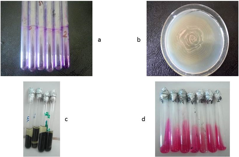
Out of the 600 collected samples, ninety-five (95, 15.83%) samples were identified as P. mirabilis isolates. P. mirabilis was detected in samples based on morphological culture’s characteristics on MacConkey agar and through microscopic examination (short bacilli and negative to gram staining), as well as some other biochemical tests mentioned in Table 2 and depicted in Figure 1. The findings about the frequency of P. mirabilis are similar to the recent report of Khan et al. [19], who reported Proteus species in pus samples with a frequency of 11% and 5.9% in urine samples. These findings are also in line with the previous study by Jabur et al. [20]. The study reported a 30% frequency of P. mirabilis in clinical samples. According to the current findings, phenotypically all P. mirabilis expressed 100% urease, 90.52% biofilm, and 92.63% swarming behaviors (Figure 1). These results strongly agree with the previous findings of Abd Al- Mayahi [16] and Sanches et al. [21].
Figure 1. Phenotypic Studies of P. mirabilis, a) Biofilm Formation, b) Swarming, c) Indole and TSI, d) Urease
3.2. Molecular Screening of P. mirabilis Virulence Factors Gene
3.2.1. ureC1 Gene Screening. The ureC1 gene-linked primer involves urease synthesis and is amplified as a 533bp fragment. In all P. mirabilis isolates, ureC1 gene showed the highest (100%) frequency as compared to other selected markers (Figure 2, Table 3). These results are similar to the previous studies of Al-Duliami et al. [22] and AL-Jumaa et al. [23].

Figure 2. Visualization of ureC1 Gene PCR Products [M= molecular ladder 100bp, Lane 1-11=isolates]
3.2.2. luxS Gene Screening. In 88 samples, a primer associated with the luxS gene, which is involved in swarming and amplified as a 464bp fragment, was found (92.63%) (Figure 3, Table 3). This finding is remarkably identical to that of Badi et al. [24], who found luxS in 70% of isolates. These findings are also consistent with those of Abd Al-Mayahi [16], who discovered that all P. mirabilis isolates (100%) display swarming behavior.

Figure 3. Visualization of PCR Products of luxS gene [M= molecular ladder 100bp, Lane 1-12=isolates]
3.2.3. rsbA Gene Screening. In 86 (90.52%) isolates, this biofilm-forming gene was found. This gene is amplified as a 467bp fragment. The rsbA gene is one of the most essential genes involved in biofilm formation and a distinguishing feature of P. mirabilis. In the current investigation, 90.52% of the tested isolates were positive for the rsbA gene (Figure 4, Table 3). Ahmadi et al. [25], revealed that 70% of the samples tested positive for rsbA gene.

Figure 4. Visualization of rsbA gene PCR Products [M= molecular ladder 100bp, Lane 1-11=isolates]
Table 3. Phenotypic and Genotypic Characteristics of Virulence Factors of P. mirabilis
|
Virulence factors |
Phenotypic n (%) |
Genotypic |
|
|
Gene name |
n (%) |
||
|
Urease production |
95 (100%) |
ureC1 |
95 (100%) |
|
Biofilm formation |
86 (90.52%) |
rsbA |
86 (90.52%) |
|
Swarming |
88 (92.63%) |
luxS |
88 (92.63%) |
The current study concludes that P. mirabilis was present in 15.83% of clinical samples in the study area. Urease production, biofilm formation, swarming virulence factors, and phenotype and genotype characteristics are present in P. mirabilis with a frequency of 100%, 90.52%, and 92.63%, respectively. This study helps in improving the knowledge of infectious behavior and provides new approaches for prevention and treatment regimes.