Detection of DNA Damage in Fish using Comet Assay
Ahmad Manan Mustafa Chatha1, Saima Naz2, Syeda Saira Iqbal3*, Azka Kirna2, Maria Lateef2, Unba Zahra2, Fatima yasmin2, Nimra Amjad2, and Ammara Javaid2
1Department of Entomology, Faculty of Agriculture and Environment, Islamia University of Bahawalpur, Pakistan
2Department of Zoology, Government Sadiq College University, Bahawalpur, Pakistan
3University of Management and Technology, Lahore, Pakistan
Abstract
Heavy metals have an enduring presence, risky characteristics, and the propensity to accumulate in the environment. This is why heavy metal toxics are widely acknowledged as harmful environmental pollutants. Heavy metals damage both aquatic and terrestrial ecosystems, posing a major risk to the environment and human health. Four freshwater fish species namely Labeo rohita, Catla catla, Hypophthalmichthys molitrix, and Ctenopharyngodon idella were the focus of this investigation. This study investigated the potential genotoxic effects of lead (Pb), copper (Cu), and cadmium (Cd) on the above fish species through the application of comet assay test. The fish were exposed to these metals at four distinct concentrations (19%, 24%, 31%, and 50% of the LC50) over the course of 40 days. All four fish species were exposed to metals to varying degrees, according to the genetic damage index, cumulative tail length of comets, and the proportion of damaged cells. In contrast to Catla catla, Hypophthalmichthys molitrix had the highest prevalence of DNA damage. The current study suggests that the presence of these particular metals in Pakistan's aquatic ecosystems may have an adverse effect on the DNA of the country's fish species. Metals cause damage to DNA in fibroblast cells through distinct mechanisms when present in water, air, and soil. Comet assay test has a remarkable sensitivity that helps to identify extremely low amounts of DNA damage. Out of the four fish species, Ctenopharyngodon idella showed higher levels of damaged cells, a higher genetic damage index, and a cumulative comet tail length as compared to others. All four fish species experienced a significant increase in DNA damage, genetic damage index, and comet tail length at 50% concentration of metals LC50.
Introduction
Aquatic creatures fulfil a significant number of nutritional demands. However, the release of industrial, agricultural, and municipal waste into aquatic environments contributes to pollution. As a result of this pollution, the consumption of fish from contaminated water sources has led to a number of health problems
[1]. Due to their extensive dispersion and notable tendency to accumulate in aquatic creatures, heavy metals play a key role in water contamination, which is primarily caused by human activities [2]. Keeping in view their negative effects on the environment, specifically the deposition and negative effects of metals on water bodies, fish, and ultimately human health, researchers have concentrated on water pollution [3–5]. The accumulation of excessive sediment in water poses a significant ecological threat as it can spread throughout the food chain. Metals have the potential to harm DNA in multiple ways and can cause damage to DNA in fibroblast cells through distinct mechanisms when present in water, air, and soil [6]. Consuming aquatic creatures contaminated with harmful heavy metals, such as fish, prawns, and shellfish, can result in the accumulation of these metals in the body's organs and chronic poisoning that may impair normal internal functioning [7].
The toxic effects of heavy metals on fish have attracted a lot of attention [8]. Our natural surroundings are rich in zinc, which is an essential micronutrient for both people and aquatic life [9, 10]. However, its excessive quantity can negatively affect living beings if it exceeds the ideal range [11]. The second essential trace element (zinc) is necessary for a number of bodily processes, including energy metabolism, bone health maintenance, protein synthesis, gene regulation, and cell membrane integrity. Over 200 enzymes, including alkaline phosphatase, carbonic anhydrase, and alcohol dehydrogenase depend on it as a co-factor in human body [12].
Environmental contaminants, such as herbicides, heavy metals, and insecticides, are known to modulate antioxidant defensive systems and to cause oxidative damage in aquatic organisms by ROS production. Several studies [13, 14] have reported that ROS generated by the metabolism of herbicides could interact with the DNA of exposed fish, resulting in the lesions detected by the comet assay. A study [15] reported that besides ROS-dependent processes, organophosphate pesticides (monocrotophos) can cause DNA strand breaks by inhibiting enzymes involved in DNA repair or by interacting with DNA.
A range of assessments are used to evaluate the genotoxic impact of metals on water pollution, which is a major concern, including in vivo biomarker assays, in vitro biomarker assays, and comet assays. Comet assay has recently received greater attention due to its remarkable sensitivity that helps to identify extremely low amounts of DNA damage, such as 0.1 DNA break per 109 Daltons [16, 17]. Reportedly, environmental monitoring has been used to assess DNA damage in individual cells [18]. A pilot study was conducted at lab scale to assess genotoxic effects [19]. Comet assay is extremely valuable for clinical research and human biomonitoring, especially since it is not always possible to collect blood or tissue samples from human subjects. In these circumstances, the comet test is used to evaluate alternative non-invasive cellular sources, such as sperm and epithelial cells [20]. Metal levels have been rising in the aquatic environment of freshwater fish, which are frequently consumed by people. Congenital and genetic abnormalities and diseases have thus increased. Sensitive genetic markers must be used to monitor the accumulation of these metals in order to stop potential disease outbreaks. Hence, the current study that uses the comet assay test was carried out to evaluate the genotoxic effects of four heavy metals on four common freshwater fish species (Labeo rohita, Catla catla, Hypophthalmichthys molitrix, and Ctenopharyngodon idella).
2. MATERIAL AND METHODS
The current study aimed to investigate the genotoxic effects of lead, copper, and cadmium on four different species of fish raised in a fish hatchery for 150 days. The fish were given two weeks to become acclimated to the lab setting before the experiment and their diet and living circumstances were closely monitored to reduce stress. Analytical grade (99.99%) lead (Pb), copper (Cu-IV), and cadmium (Cd) compounds were used to make stock solutions for genotoxicity which were diluted to the desired metal concentrations. Probit analysis, a statistical technique used to determine the concentration of a substance that is lethal to 50% of the test population, was used to calculate the 96-hour LC50 of each metal for the fish [21].The same water (30o C), pH (7.5), and hardness (300 mg/L-1) were maintained for all tests. Each fish species was individually treated with four different sub-lethal water-borne dosages to assess DNA damage in erythrocytes. Pb, Cu, and Cd were exposed to concentrations that were 19%, 24%, 31%, and 50% of LC50. For the duration of the trial, the fish were fed twice daily until they were satisfied with a meal that contained 3 Kcal/g of calories and 34% DE (Digestible Energy).
Blood samples were collected from the caudal vein of the fish. These samples were then processed in accordance with the guidelines for the comet assay test [22]. Using 0.4 M Tris buffer with a pH of 7.5, the slides were sensitively neutralized after electrophoresis and the DNA was then stained with ethidium bromide (20 g/ml). A low-light camera and an epi-fluorescence microscope with mercury short arc reflector lamp filters for ethidium bromide at 400 X magnification were used to examine 150 cells (50 cells per replication) at random. DNA-damaged cells looked like comets, although cells without DNA damage had a nucleus without a tail. The extent of DNA damage was calculated using the length of DNA movement in the comet tail [23]. Since they were classified as apoptotic cells, cells with no head or a dispersed head were excluded from the investigation. Visually classifying cells into five groups or ‘comets’ according to the length of their tails allowed the researchers to gauge the extent of DNA damage. Type 0 refers to unharmed cells, Type I to cells with low level of damage, Type II to cells with medium level of damage, Type III to cells with high level of damage, and Type IV to totally damaged cells. In order to analyse statistical differences between means, Newnan-Keul test was utilized and both parametric and non-parametric tests were performed to evaluate differences at a significance level of p > 0.05. Metal concentrations in the fish's gills, liver, kidney, fins, bones, muscles, and skin were determined. Measurements were made both before and after the fish were exposed to 19 various combinations. Acute toxicity tests were carried out in accordance with the recommended procedures [24] to figure out the lethal and 96-hour LC50 concentrations.
3. RESULTS
Table 1 shows that Labeo rohita, Hypophthalmichthys molitrix, Catla catla, and Ctenopharyngodon idella all had significantly different 96-hour LC50 values for Pb, Cu, and Cd. Table 2 lists the effects of four distinct metal concentrations on the four fish species' peripheral blood erythrocytes in terms of cell damage, genetic damage index, cumulative comet tail length, and negative and positive control treatments. All four fish species experienced a significant increase in DNA damage, genetic damage index, and comet tail length at 50% of metals LC50. The only exception was Ctenopharyngodon idella which showed a higher level of damaged cells, a higher genetic damage index, and cumulative comet tail length as a result of exposure to the positive control treatment rather than Cu. Moreover, peripheral blood erythrocytes from all three metals exhibited a concentration-dependent rise in DNA damage in the four different fish species (Table 2). The results of the current study on the effectiveness of the four fish species are presented in Table 3.
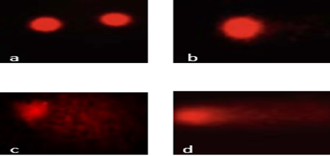
Figure 1. Comet Assay Showing DNA Damage in (a) Labeo rohita, (b) Hypophthalmichthys molitrix, (c) Catla catla, and (d) Ctenopharyngodon idella
Table 1. Acute Toxicity (96-hr LC50 and Lethal Concentrations) of Metals
|
Fish |
Metal |
Mean 96-hr LC50(µgL-1) |
95 % C.I.(µgL-1) |
Mean Sub-lethal Concentrations (µgL-1) |
95 % C.I.(µgL-1) |
|
Fish 1 |
Pb |
59.26±0.61 |
50.02-65.81 |
104.66±0.53 |
93.87-125.28 |
|
Fish 2 |
78.75±0.56 |
70.62-85.69 |
128.53±0.69 |
116.86-148.46 |
|
|
Fish 3 |
74.13±0.57 |
66.77-80.19 |
117.69±0.55 |
17.81-134.41 |
|
|
Fish 4 |
61.17±0.38 |
55.22-66.51 |
100.08±0.56 |
90.77-115.98 |
|
|
Fish 1 |
Cd |
78.24±0.39 |
71.40-84.22 |
118.63±0.74 |
108.34-137.23 |
|
Fish 2 |
69.89±0.94 |
63.36-75.73 |
109.78±0.87 |
100.36-125.27 |
|
|
Fish 3 |
65.24±0.24 |
57.59-71.81 |
113.38±0.20 |
101.63-134.35 |
|
|
Fish 4 |
50.38±0.36 |
44.92-55.05 |
81.41±0.42 |
73.65-95.24 |
|
|
Fish 1 |
Cu |
85.96±0.45 |
79.33-91.60 |
122.49±0.60 |
113.85-136.85 |
|
Fish 2 |
75.95±0.45 |
69.67-81.11 |
108.94±0.36 |
100.66-123.89 |
|
|
Fish 3 |
63.17±0.33 |
55.91-68.87 |
99.44±0.41 |
90.12-117.16 |
|
|
Fish 4 |
62.91±0.74 |
55.11-69.18 |
102.45±0.64 |
92.08-122.25 |
Figure 2. Acute Toxicity (96-hr LC50 and lethal concentrations) of Metals
Table 2. DNA Damage in Peripheral Erythrocytes of Fish Due to Metal Exposure
|
Species |
Parameters |
Exposure Concentrations (mg L-1) |
%age of damaged cells (II+III+IV) |
Genetic Damage Index *(GDI) |
Cumulative Tail Length (µm) |
|
Fish 1 |
Control |
0.00 |
3±3f |
0.06±0f |
3±0f |
|
Positive control (+) |
CP (20 µgg-1) |
34±4d |
2.3±0d |
131±0d |
|
|
19% of LC50 |
20.38 |
15±5e |
0.8±0e |
38±0e |
|
|
24% of LC50 |
36.27 |
98±2c |
3.7±0c |
145±0c |
|
|
31% of LC50 |
45.46 |
85±3b |
3.2±0b |
175±0b |
|
|
50% of LC50 |
6.48 |
87±4a |
3.5±0a |
286±1a |
|
|
Fish 2 |
Negative control (-) |
0.00 |
1±1.1e |
0.1±0.0c |
3±0.f |
|
Positive control (+) |
CP (20 µgg-1) |
35±1.1a |
3.6±0.0a |
90±0.2b |
|
|
17% of LC50 |
8.03 |
11±2.5d |
0.6±0.0d |
52±0.2e |
|
|
25% of LC50 |
12.54 |
15±4.0c |
±0.0c |
92±1.8d |
|
|
33% of LC50 |
23.78 |
49±0. b |
2.0±0.0b |
87±0.3c |
|
|
50% of LC50 |
32.81 |
57±0.0a |
2.7±0.0a |
111±0.4a |
|
|
Fish 3 |
Negative control (-) |
0.00 |
2±3f |
0.06±0f |
4±0f |
|
Positive control (+) |
CP (20 µgg-1) |
36±4d |
1.3±0d |
131±0d |
|
|
19% of LC50 |
19.38 |
14±5e |
0.5±0e |
39±0e |
|
|
24% of LC50 |
35.27 |
97±2c |
3.6±0c |
146±0c |
|
|
31% of LC50 |
45.46 |
86±3b |
3.5±0b |
174±0b |
|
|
50% of LC50 |
62.48 |
89±4a |
3.8±0a |
288±1a |
|
|
Fish 4 |
Negative control (-) |
0.00 |
1±1.1e |
0.2±0.0c |
2±0.f |
|
Positive control (+) |
CP (20 µgg-1) |
39±1.1a |
3.5±0.0a |
89±0.2b |
|
|
19% of LC50 |
8.03 |
14±2.5d |
0.5±0.0d |
51±0.2e |
|
|
24% of LC50 |
12.54 |
15±4.0c |
9.2±0.0c |
93±1.8d |
|
|
31% of LC50 |
23.78 |
49±0. b |
1.0±0.0b |
88±0.3c |
|
|
50% of LC50 |
32.81 |
57±0.0a |
2.6±0.0a |
115±0.4a |
Note. The various lowercase letters (a, b, c, d, e, f) show a highly significant difference in DNA damage in the peripheral erythrocytes of fish due to metal exposure.
The researchers investigated how metals affect cell DNA and cause damage in four different fish species. Following lead exposure, all four species were noticeably more likely to have damaged cells, with silver carp having the greatest percentage of damage, followed by Labeo rohita, Ctenopharyngodon idella, and Catla catla. Lead exposure considerably increased the sensitivity of all four species as compared to copper exposure, which led to the minimum number of injured cells. The order of metal toxicity responsible for damaging the erythrocytes of the four fish species was Pb (1.4±0.5)> Cu (1.2±0.7) > Cd (1.1±0.3). The values of the fish's genetic damage index significantly varied as a result of their exposure to the metals. The highest damage was caused by lead exposure, closely followed by cadmium and copper exposures. Grass carp showed greater damage from lead exposure and lesser damage from copper exposure, silver carp showed the highest mean value due to lead exposure, followed by cadmium. Lead substantially harmed the erythrocytes of all four fish species. The species most vulnerable to induced DNA damage was Hypophthalmichthys molitrix, followed by Labeo rohita, Ctenopharyngodon idella, and Catla catla. All four species were more susceptible to lead exposure than to the other elements. Catla catla and Ctenopharyngodon idella were least susceptible to cadmium exposure, while Labeo rohita and Hypophthalmichthys molitrix were least susceptible to copper exposure. The most sensitive of the four species was the silver carp, which was then followed by Labeo rohita, Catla catla, and Ctenopharyngodon idella. The average susceptibility of the four species to the three metals differed greatly. Table 3 provides the details.
3.1. Bioaccumulation of Metals in Fish Organs
According to the quoted study, the metals were discovered to rise dramatically in the bodies of fish when exposed to a quantity that resulted in a 96-hour LC50. Labeo rohita showed a stronger propensity to accumulate lead, while Catla catla demonstrated a stronger propensity to accumulate copper and cadmium. The fish's gills tended to accumulate cadmium more frequently, while the liver of the fish accumulated considerable levels of lead and copper. Lead, cadmium, and copper were the metals with the highest concentrations in the fish's bodies after exposure.
Table 3. DNA Damage (%) in Fish under Exposure to Heavy Metals
|
Fish |
Exposure Metal |
||
|
Lead |
Cooper |
Cadmium |
|
|
Percentage of damaged cells (%) |
|||
|
Fish 1 |
45±29 |
50±5 |
40±32 |
|
Fish 2 |
29±15 |
39±8 |
55±40 b |
|
Fish 3 |
40±29 |
49±5 |
49±33b |
|
Fish 4 |
28±15 |
39±8 |
56±39 b |
|
*Genetic Damage Index (GDI) |
|||
|
Fish 1 |
1.3±0.7 |
1.2±0.7 |
1.3±0.1 |
|
Fish 2 |
1.2±0.6 |
1.3±0.6 |
1.2±0.2 |
|
Fish 3 |
1.4±0.5 |
1.1±0.4 |
1.4±0.4 |
|
Fish 4 |
1.1±0.4 |
1.4±0.5 |
1.1±0.3s |
|
Cumulative Tail Length (µm) |
|||
|
Fish 1 |
112±67 |
104±75 |
97±80 |
|
Fish 2 |
114±68 |
102±73 |
98±81 |
|
Fish 3 |
113±65 |
103±74 |
96±79 |
|
Fish 4 |
111±64 |
105±76 |
95±82 |
Table 4. Bioaccumulation of Metals (µgg-1) in Fish Organs
|
Metals in Fish Species |
Organs |
||||||||
|
Kidney |
Liver |
Skin |
Muscle |
Fins |
Gills |
Bones |
Means |
||
|
Pb |
Fish 1 |
76.58±2.21 d |
432.88±1.32 d |
150.90±1.71c |
122.59±1.41cd |
309.35±1.25b |
233.96±2.24 b |
68.84±0.92bc |
199.30±134.27d |
|
Fish 2 |
407.57±3.55 b |
502.50±4.69 b |
120.58±0.52d |
155.50±0.84 b |
148.03±3.54 d |
467.10±3.22 a |
71.78±1.45bc |
267.58±183.27a |
|
|
Fish 3 |
303.95±3.64 c |
60.98±4.68 e |
310.05±2.41a |
114.17±3.62 d |
39.91±0.28 e |
91.27±2.65 e |
62.79±1.75 c |
140.45±116.24c |
|
|
Fish 4 |
550.00±1.32 a |
454.17±2.75 c |
53.34±0.98 e |
22.59±1.05 e |
456.29±2.11 a |
152.06±1.13 c |
73.31±1.13bc |
251.68±225.61b |
|
|
Cu |
Fish 1 |
17.14±0.94 de |
178.18±1.64 a |
186.64±4.19a |
16.24±1.74 d |
345.46±3.22 a |
311.83±3.88 b |
34.76±1.85 b |
155.75±138.53a |
|
Fish 2 |
485.00±2.50 a |
31.00±0.50 e |
43.22±2.61 d |
28.27±0.48 c |
130.88±5.89 c |
259.11±3.62 d |
21.93±2.31d |
142.77±173.63b |
|
|
Fish 3 |
14.68±3.34 e |
62.50±1.29 c |
70.60±0.41 c |
57.25±1.21 b |
47.36±0.59 e |
31.55±2.08 e |
26.68±0.40c |
44.37±20.61 c |
|
|
Fish 4 |
47.59±1.16 b |
85.09±2.01 b |
17.73±0.49 e |
15.69±1.81 d |
221.34±2.03 b |
287.18±0.79 c |
15.34±3.79 e |
98.57±110.86 d |
|
|
|
Fish 1 |
16.11±0.71 c |
106.21±2.51 a |
48.34±2.84 a |
17.07±0.39 a |
77.41±1.13 a |
9.53±0.42 e |
8.28±0.39 de |
40.42±38.44 a |
|
Cd |
Fish 2 |
12.24±0.92 de |
10.04±0.78 e |
19.94±0.80 b |
8.56±0.80 b |
32.54±2.59 b |
86.55±1.25 a |
15.89±2.52 c |
26.54±27.64 b |
|
|
Fish 3 |
16.70±0.73 bc |
16.74±0.66 b |
16.19±1.43 c |
6.02±0.20 cd |
5.08±0.78 e |
19.40±2.63 c |
21.92±1.74ab |
14.58±6.44 c |
|
|
Fish 4 |
11.07±0.65 e |
10.77±0.31 de |
6.72±0.36 d |
4.42±0.47 d |
14.18±1.45 c |
23.40±0.62 b |
7.15±0.29 e |
11.10±6.32d |
Note. FISH 1 = Catla catla; FISH 2 = Labeo rohita; FISH 3 = Ctenopharyngodon idella; FISH 4 = Hypophthalmichthys molitri
4. DISCUSSION
The use of chemicals in agriculture and other industries has led to an increase in genetic abnormalities, diseases, and mortality rates. Exposure to these substances by creatures in natural settings causes these problems [25].The effects of chemicals on the composition and functionality of cellular DNA requires further study. In this regard, it has been demonstrated that an increase in metallic ion concentration is also accompanied by an increase in DNA damage in two separate peripatetic fish species. Similarly, when exposed to varied levels of arsenic, DNA damage in the blood cells of Oreochromis mossambicus significantly increased in comparison to the control group [26]. The current study's two fish species showed a significant capacity to incur harm to their DNA when exposed to Zn. Furthermore, previous research demonstrated that the DNA of Clarias gariepinus is adversely affected by metals, such as Zn, Cu, As, Pb, and Cd [2]. Metals affect enzymes that prevent oxidation by functioning as antioxidants. Fish experience oxidative effects as a result of the increase in lipid and glutathione peroxidation. Additionally, it has been noted that increased aluminum exposure causes Ctenopharyngodon idella to gradually produce more superoxide dismutase, which helps to counteract the negative effects of reactive oxygen species [27]. The comparative analysis of DNA damage by comet assay and chromosome damage by FISH-comet assay in the same samples may be useful to improve the interpretation of the results of FISH-Comet assay (with the application of whole and partial chromosome probes).
In the only work of this kind [28], it was shown that chromosome damage in human blood cells treated with doxorubicin, as measured in FISH studies (using chromosome 1 probe), appeared to be a more sensitive endpoint than strand breaks in the comet assay, since chromosomal damage was observed at lower doses (e.g. 1.25 μM vs. 0.1 μM). Very high correspondence was found between the results obtained in the chromosomal aberration test and the comet assay using human peripheral lymphocytes and Chinese hamster lung cells treated with methyl methanesulfonate, cyclophosphamide, and novel pharmaceuticals. However, positive results in the comet assay were always observed at higher doses than those that produced an effect in the cytogenetic test [29]. The results of this study are also in line with another study which found that the pathogenicity of Al, Cu, Cr, Ni, Zn, Fe, and Pb caused more DNA damage to peripheral red blood cells (RBCs) of the domestic fish species Hyphssobryconluetkenii [30]. Carassius auratus and Channa punctatus (which are vertebrates) are the best choice to estimate possible dangers because of their capacity to metabolize and collect contaminants in their bodies [31]. Peripheral blood provides an organism's complete health status. RBCs are, therefore, ideal for analysing DNA damage. They are suitable and nucleated, so blood cells attracted attention from the adversary nucleoidselectrophoresis of isolated gels [32]. In this experiment, Hypophthalmichthys molitrix had a higher genetic damage index than Labeo rohita, Catenophryngodon idella, and Catla catla. Pb>Cu>Cd is the order in which different metals damaged DNA in terms of the cumulative length of comet tails [33]. Comets in Cobitis expanded RBCs have longer cumulative tail lengths because of the pathogenicity of industrial pollution that contains As, Cu, Hg, Cr, and Mn. Due to a higher amount of tail DNA than the other three fish in the study, Hypophthalmichthys molitrix displayed a higher susceptibility to metals.
Due to the differences in their physiology and species-selectivity to interchange against various metals, all species exhibited distinct responses to each metal as well as to the mixture of metals. Metals harm DNA and it raises serious concerns about their potential threat to carp survival in natural aquatic environments. According to [34], the amount of metals ingested, how they are distributed throughout the body, and the quantity in which they are retained in various tissues determine the amount of metals that accumulate in an organism. According to a study, fish can modify their carriers to adapt to high zinc concentrations in water [35]. The buildup of metals can become dangerous for both people and the creatures that come into contact with them. In order to accurately detect environmental pollution, it is possible to look at the pattern of metal buildup in fish tissues. The three most obvious sites of metal accumulation in fish are the liver, kidneys, and gills, with the liver showing the highest amount of persistence [6]. The significantly higher metal levels in mixture-stressed fish can be explained by the binding of metals to metallothionein in the liver and other proteins involved in metal metabolism [36].
4.1. Conclusion
All four fish species displayed various degrees of genotoxic damage in their peripheral blood erythrocytes, according to the comet assay utilised in the current study. In order to monitor metallic ion pollution in natural aquatic ecosystems, the findings suggest using Labeo rohita, Hypophthalmichthys molitrix, Catla catla, and Ctenopharyngodon idella as suitable bioindicators.
Conflict of Interest
The author of the manuscript has no financial or non-financial conflict of interest in the subject matter or materials discussed in this manuscript.
Data Availability Statement
The data associated with this study will be provided by the corresponding author upon request.
Bibliography
- Pradeepkiran JA. Aquaculture role in global food security with nutritional value: a review. Translat An Sc. 2019;3(2):903–910. https://doi.org/10.1093/tas/txz012
- Farombi EO, Adelowo OA, Ajimoko YR. Biomarkers of oxidative stress and heavy metal levels as indicators of environmental pollution in African cat fish (Clarias gariepinus) from Nigeria Ogun River. Int J Environ Res Public Health. 2007;4(2):158–65. https://doi.org/10.3390/ijerph2007040011
- Mudgal V, Madaan N, Mudgal A, Singh RB, Mishra S. Effect of toxic metals on human health. Open Nutraceut J. 2010;3:94–99. https://doi.org/10.2174/18763960010030100094
- Naz S, Mansouri B, Chatha AM, et al. Water quality and health risk assessment of trace elements in surface water at Punjnad Headworks, Punjab, Pakistan. Environ Sci Pollut Res. 2022;29(40):61457–61469. https://doi.org/10.1007/s11356-022-20210-4
- Kakade A, Sharma M, Salama ES, et al. Heavy metals (HMs) pollution in the aquatic environment: Role of probiotics and gut microbiota in HMs remediation. Environ Res. 2023;223:e115186. https://doi.org/10.1016/j.envres.2022.115186
- Naz S, Javed M. Growth responses of fish during chronic exposure of metal mixture under laboratory conditions. Pak Vet J. 2013;33(3):354–357.
- Zhou X, Sun J, Zhu G, Ge C. Toxicity of copper, zinc, lead, cadmium to tissue's cellular DNA of the fish (Carassius auratus). Acta Agricul Nucl Sinica. 2001;15(3):167–73.
- Yang J, Yin J, Xu HL, Gong XQ, Yu RP, Wang D. The ingestion and accumulation of zinc, copper, lead, cadmium and arsenic in the Yangtze finless porpoise, Neophocaena phocaenoides asiaeorientalis. Acta Hydrobiol Sinica. 2005;29(5):667–563.
- Wang WN, Liang H, Wang AL, Chen T, Zhang SE, Sun RY. Effect of pH and Zn(2+) on subcultured muscle cells from Macrobrachium nipponense. Methods Cell Sci. 2000:22(4):277–284. https://doi.org/10.1023/A:1017962429862
- Isangedighi IA, David GS. Heavy metals contamination in fish: effects on human health. Journal of Aquatic Science and Marine Biology. J Aquatic Sci Marine Biol. 2019;2(4):7–12.
- Chu EW, Karr JR. Environmental impact: concept, consequences, measurement. In: Roitberg BD, ed. Reference Module in Life Sciences. Elsevier; 2017. https://doi.org/10.1016/B978-0-12-809633-8.02380-3
- Dawood MA, Alagawany M, Sewilam H. The role of zinc microelement in aquaculture: a review. Biolog Trace Elem Res. 2022;200(8):3841–3853. https://doi.org/10.1007/s12011-021-02958-x
- Liu Y, Zhang Y, Liu J, Huang D. The role of reactive oxygen species in the herbicide acetochlor-induced DNA damage on Bufo raddei tadpole liver. Aqu Toxicol. 2006;78(1):21–26. https://doi.org/10.1016/j.aquatox.2006.01.016
- Cavalcante DG, Martinez CB, Sofia SH. Genotoxic effects of Roundup® on the fish Prochilodus lineatus. Mutat Res Rev Mutat Res. 2008;655(1-2):41–46. https://doi.org/10.1016/j.mrgentox.2008.06.010
- Thakur S, Sarkar B, Dhiman M, Mantha AK. Organophosphate-pesticides induced survival mechanisms and APE1-mediated Nrf2 regulation in non-small-cell lung cancer cells. J Biochem Molecul Toxicol. 2021;35(2):e22640. https://doi.org/10.1002/jbt.22640
- Ali F, El-Shehawi AM, Seehy MA. Micronucleus test in fish genome: a sensitive monitor for aquatic pollution. Afr J Biotechnol. 2008;7(5):606–612.
- Al-Saeed FA, Naz S, Saeed MH, et al. Oxidative stress, antioxidant enzymes, genotoxicity and histopathological profile in oreochromis niloticus exposed to lufenuron. Pak Vet J. 2023;43(1):160–166. https://doi.org/10.29261/pakvetj/2023.012
- de Andrade VM, de Freitas TR, da Silva J. Comet assay using mullet (Mugil sp.) and sea catfish (Netuma sp.) erythrocytes for the detection of genotoxic pollutants in aquatic environment. Mut Res Gene Toxicol Environ Mutag. 2004;560(1):57–67. https://doi.org/10.1016/j.mrgentox.2004.02.006
- Emmanouil C, Smart DJ, Hodges NJ, Chipman JK. Oxidative damage produced by Cr (VI) and repair in mussel (Mytilus edulis L.) gill. Mar Environ Res. 2006;62:S292–S296. https://doi.org/10.1016/j.marenvres.2006.04.024
- Rojas E, Lorenzo Y, Haug K, Nicolaissen B, Valverde M. Epithelial cells as alternative human biomatrices for comet assay. Front Gnetics. 2014;5:e386. https://doi.org/10.3389/fgene.2014.00386
- Azmat H, Javed M, Jabeen G. Acute toxicity of aluminium to the fish (Catla catla, Labeo rohita and Cirrhina mrigala). Pak Vet J. 2012;32(1):85–87.
- Singh NP, McCoy MT, Tice RR, Schneider EL. A simple technique for quantitation of low levels of DNA damage in individual cells. Experiment Cell Res. 1988;175(1):184–191. https://doi.org/10.1016/0014-4827(88)90265-0
- Grover P, Danadevi K, Mahboob M, Rozati R, Banu BS, Rahman MF. Evaluation of genetic damage in workers employed in pesticide production utilizing the Comet assay. Mutagenesis. 2003;18(2):201–205. https://doi.org/10.1093/mutage/18.2.201
- Rice EW, Bridgewater L, American Public Health Association. Standard Methods For The Examination of Water And Wastewater (Vol. 10). Washington, DC: American Public Health Association; 2012.
- Livingstone DR. Contaminant-stimulated reactive oxygen species production and oxidative damage in aquatic organisms. Marine Pollut Bullet. 2001;42(8):656–666. https://doi.org/10.1016/S0025-326X(01)00060-1
- Ahmed MK, Habibullah-Al-Mamun M, Hossain MA, et al. Assessing the genotoxic potentials of arsenic in tilapia (Oreochromis mossambicus) using alkaline comet assay and micronucleus test. Chemosphere. 2011;84(1):143–149. https://doi.org/10.1016/j.chemosphere.2011.02.025
- Van der Oost R, Beyer J, Vermeulen NP. Fish bioaccumulation and biomarkers in environmental risk assessment: a review. Environ Toxicol Pharmacol. 2003;13(2):57–149. https://doi.org/10.1016/S1382-6689(02)00126-6
- Spivak G. The Comet-FISH assay for the analysis of DNA damage and repair. Fluorescence in situ Hybridization (FISH) protocols and applications. Meth Molecul Biol. 2010;659:129–145.
- Johannes C, Obe G. Chromosomal aberration test in human lymphocytes. Methods Molecul Biol. 2019;2031:121–134.
- Scalon MC, Rechenmacher C, Siebel AM, et al. Evaluation of sinos river water genotoxicity using the comet assay in fish. Braz J Biol. 2010;70:1217–1222.
- Filice M, Cerra MC, Imbrogno S. The goldfish Carassius auratus: an emerging animal model for comparative cardiac research. J Compar Physiol B. 2022;192(1):27–48. https://doi.org/10.1007/s00360-021-01402-9
- Costa PM, Neuparth TS, Caeiro S, et al. Assessment of the genotoxic potential of contaminated estuarine sediments in fish peripheral blood: laboratory versus in situ studies. Environ Res. 2011;111(1):25–36. https://doi.org/10.1016/j.envres.2010.09.011
- Marčić MM, DoleNec Z, Mrakovčić M. Assessment of DNA integrity in erythrocytes of Cobitis elongata affected by water pollution: the alkaline comet assay study. Folia Zoolog. 2008;57(1-2):120–130.
- Naz S, Chatha AM. Metals mixture effects on growth performance and their bioaccumulation in fish. Iran J Fish Sci. 2022;21(2):605–618.
- Naz S, Hussain R, Ullah Q, Chatha AM, Shaheen A, Khan RU. Toxic effect of some heavy metals on hematology and histopathology of major carp (Catla catla). Environ Sci Pollution Res. 2021;28(6):6533–6539. https://doi.org/10.1007/s11356-020-10980-0
- Naz S, Javed M. Evaluation of acute toxicity of metals mixture and bioaccumulation in freshwater fish. Biosci Methods. 2013;4(3):11–18. https://doi.org/ 10.5376/bm.2013.04.0003