
Muhammad Javaid Afzal1*, Farah Javaid2, Muhammad Ilyas Yasin3, Shahzadi Tayyaba4, and Muhammad Waseem Ashraf5
1Department of Physics, Govt. Islamia Graduate College Civil Lines Lahore, Pakistan
2Department of Physics, Govt. APWA Graduate College (W), Lahore, Pakistan
3Department of Firearms Acadiana Criminalistics Laboratory, New Iberia Louisiana – USA
4Department of Information Sciences Division Science and Technology, University of Education, Lahore, Pakistan
5Department of Electronics, Govt. College University, Lahore, Pakistan
* Corresponding Author: [email protected]
A microelectromechanical system (MEMS) is a diminutive machine having electronic and mechanical components with a size ranging from 20 µm-1 mm. In this present-day world, MEMS fabrication techniques have remodeled the conventional approaches towards system fabrication. Microfluidics is an eminent domain of MEMS in which small volumes of fluids are disciplined in micro-channels having dimensions in the submillimeter to achieve the desired outputs. Microfluidics have revolutionized the realm of compact system fabrication through preeminent inventions like lab-on-a-chip technology. Microchannels of various architectures are fabricated to employ microfluidic systems depending upon the required function of the device. In ophthalmology, Retinal Vein Occlusion (RVO) is an ailment in which small veins that take away blood from the human eye's retina are blocked or fissured, causing vision loss. Therefore, in this study, four micro-channels with different architectures, namely, sinusoidal, U-shaped, spiral, and curvilinear, were simulated by using the fuzzy technique to investigate the optimization of fluids for the implantation process to fix the RVO elixir. The two most critical parameters in retinal vein flow rate and velocity were taken at the output for optimization. Hence, fuzzy fluidic simulation revealed that curvilinear micro-channels were the best fit for biomedical implantation to treat RVO malady.
Keywords: biomedical implants, curvilinear microchannel, fuzzy logic, microchannels, retinal vein occlusion
A device with electrical and mechanical components as its integral parts and a size less than 1mm but greater than one micron falls in the category of MEMS [1]. Applications of MEMS have greatly revolutionized almost every field of scientific research. Additionally, MEMS based devices are widely employed in routine life; including automobile airbag accelerometers, keyless entry mechanisms, heat exchanging systems for cooling microsystems, lab-on-a-chip technology, micro drug delivery mechanisms, and many more. In the 1990s, an essential sector of MEMS, microfluidics study, came into the limelight with its astonishing applications in the fabrication of MEMS-based devices. In microfluidics systems, small volumes of fluids, usually the norm in the range of microliters (µl) to picoliters (pl) are controlled and manipulated in micro-ducts having dimensions from tens to hundreds of micrometers to obtain desired outputs [2]. Inkjet printers proved to be a prominent commercial success for microfluidics-based MEMS [3]. Microfluidics-based devices greatly benefited medical science by providing biochips, especially diagnosis applications [4].
Micro-channels are an imperative part of any microfluidic device [5]. The small volume of fluids is contained and manipulated in micro-channels of hydraulic diameter customarily below than1mm. Originally the concept of micro-channels was suggested by Tuckerman and Pease of Stanford Electronics Laboratories [6]. Micro-heat exchangers have widely employed micro-channels containing small qualities of fluids for heat exchange. These micro-heat exchangers play a significant role in the gas turbine engines, air conditioning, and heat pumps. It is because of the modest velocities at which fluids moving via microchannels, low Reynolds numbers are obtained [7]. Primarily, this paper is based on Retinal Vein Occlusion (RVO) for all three micro-channels. The human eye's lens focuses on all the incoming light onto the retina, located in the rear of the eyeball. The optical nerve transmits information from the retina to the brain, where it is processed to recognize images. Retinal vein occlusion (RVO) occurs when a clot blocks blood flow in the retinal vein. Hence, this research describes the maximal and typical blood flow rates in the human retinal veins.
Depending upon the intended purpose, micro-channels of different morphology were concocted. For example, to reinforce the flow pattern like parallel, serpentine, crossed, oblique, and ratchet morphology of micro-channels could have adopted the architecture application. The shape of the microchannel is always an important factor to obtain the required results. Computational fluid dynamics (CFD) is often employed to optimize the desired microchannel shape [8]. The design of different alphabets like J, Y, T, U, and S can be a choice for purpose-based on its applications [9]. The other viable architectures include triangular, spiral, double spiral, trapezoidal, rectangular, circular, square, coil, double coil, curved, curvilinear, concentric, and sinusoidal with ascending and descending orders. The preferred choice of shape may vary greatly depending upon the desired results. Fluids exhibit differently in different shapes of microchannel. The flow rate and speed of the fluid in the microchannel are considered most meaningful for its suitability in any application. The chemistry of blood remained the same in all micro-channels [10].
Four micro-channels were subjected for the fluid optimization in the current study, which are briefly discussed below.
A sinusoidal shape microchannel comprised of periodic repetitions of oscillations [11]. Sinusoidal microchannels of defined wavelength and amplitude have been employed in previous research for heat dissipation and mass transformation [12]. Studies have showed that sinusoidal micro-channels exhibit excellent flow conditions for the blood-like fluid. Blood flow at curves tends to lose its streamlined flow; however, regained its normal tendon after the curves. Such micro-channels have been employed for biomedical implantations for varicose veins. Such micro-channels possessed excellent qualities for dissipation of heat because of the morphology [13]. Curvatures helped to resist and streamlining the flow by making it easier to homogenize in the fluids.
Figure 1. Sinusoidal Microchannel
1.1.2. Spiral Microchannel. A coil-like microchannel called a spiral would have more complexity with increasing numbers of twists. Therefore, the current study's application demanded minimum twist in channels. Continuous centrifugal force made the blood lose its streamline flow [14]. Morphology of retinal veins can easily afford the implantation of spiral micro-channels. Blood rheology easily allowed the use of a spiral channel for implantation purposes [15]. Such micro-channels have been widely employed in mass transit systems [16].
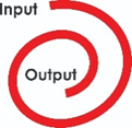
Figure 2. Spiral Microchannel
1.1.3. U-Shaped Microchannels. Researchers have been employing U-Shaped microchannels in fluid dynamics effectively [17]. A big advantage of a U-shape is its ability to adjust its length as per need [18]. The single curve in such a channel tends to offer minimum parametric changes as compared to other shapes [19]. However, Its biomedical application is well investigated in previously done research [20].
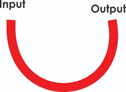
Figure 3. U-Shaped Microchannel
1.1.4. Curvilinear Microchannel. A microchannel comprising of curving boundaries or edges is called a curvilinear microchannel. Blood particles in its plasma offer the same behaviour to curvilinear morphology fabricated microchannel as that of natural response to veins [21]. More prone to extendable flexibility, its suitability for biomedical implants is favourable [22]. With the increased number of curves, the cell separation task can be obtained from such micro-channels [23].

Figure 4. Curvilinear Microchannel
Biological systems in human organs, namely, brain, liver, lungs, kidney, heart, and eyes contain many micro-channels majorly performing the critical functions of mass and heat transfer [24]. Cells can also migrate via micro-channels in a body structure [25]. It is believed that a comprehensive microchannel mass transfer mechanism work in human biological systems. Human tissues contain a complex network of micro-channels, which can be used to regulate routine tissue operations [26]. Primarily, blood composed of two major components; plasma and red blood cells. Plasma is a Newtonian fluid and red blood cells are intransigent spherical particles [27]. Blood bank viscosity is based on the percentage of blood that is made up of red blood cells. Characteristics of flowing blood like blood pressure, velocity, flow rate, the flow pattern is affected by the structure of the microchannel. Naturally occurring micro-channels in human biological system vary in morphology, which depends upon the function they are performing [28]. However, most micro-channels of human biological systems are elastic and can assume different morphology with the varying position of the human body [29]. Similarly, the diameter of this microchannel also responded to the varying volume and pressure of blood and adjusted accordingly to tolerate the instant conditions.
The lens of the human eye focuses on all coming rays to form an image on the inner side of the eye sphere to a place called the retina [30]. Nerve cells surrounding the retina convert the coming light into signals, which are transmitted to the brain via optical nerve for image recognition. Blockage of blood flow in the retinal vein because of a clot is called retinal vein occlusion (RVO). RVO is one of the most commonly occurring retinal vascular disease with an estimated 16 million effete globally [31]. A person affected by RVO may suddenly lose partial or full vision permanently without pain. So far iRVO is incurable; therefore, damage once done is irreversible but ophthalmologists can treat patients to stop further deterioration. Individuals suffering from diabetes and high blood pressure are more prone to RVO [32]. Other commonly found reasons for RVO include hypertension, high cholesterol, smoking, glaucoma, and excessive weight [33].
Furthermore, RVO is additionally categorized into two types; Central Retinal Vein Occlusion (CRVO) and Branch Retinal Vein Occlusion (BRVO). In CRVO the main retinal vein carrying blood is blocked/raptured, whereas BRVO affects a small branch of the main retinal vein. Both central and branch retinal veins have diameters in the range of µm with the central vein having a larger diameter as compared to the other branches. In certain cases, blocked vessels caused the leakage of jelly-like fluid into the retina resulting in small spots. These spots hampered the transmission of optical signals, which can be further used for the formation of an image. In other cases, some new unusual blood vessels may be caused by the retina; a process called neovascularization. These newly developed vessels leak the blood or fluid giving rise to RVO [34].
Different imaging tests are used to see the footsteps of RVO in the human eye, which include fluorescein Angiography, Optical Coherence Tomography, and Fundus Autofluorescence [35]. In retinal veins of human's maximum blood velocity and average flow rate as 54 mm/s and 2.04 nanoliters/ sec, respectively. Retinal veins carry blood away from the retina are quite similar to the microchannels.
1.3.1. Current Treatments. Current ophthalmology techniques to treat RVO include anti-vascular endothelial growth factor drug injections, retina opening massage, clot-busting medication, steroid treatment, Panretinal photocoagulation, focal laser treatment, and hyperbaric oxygen therapy [36]. These treatments deal with the issues arising from RVO to prevent further damage.
1.3.2. Challenges. There is no treatment or medication, which can completely unblock retinal veins [35]. Until now, RVO is incurable; ophthalmologists only manage the damage and prevent further deterioration. Vision problems, such as blurring or complete loss of sight in one eye, may appear unexpectedly. Complications from RVO include ocular edoema and haemorrhage. These conditions can cause blindness if not treated at the right time.
Biomedical implants referred to placing natural or fabricated devices or tissues inside or on the surface of the human body to accomplish specific tasks. There may be different reasons for biomedical implants, which may include a missing body organ for supplemental support and monitoring body functions [37]. Hence, the implanted devices or tissues may be removed when they are no longer needed for the process. Primarily, operational surgery was required for the implantation. Most commonly implanted devices included implantable cardioverter defibrillators (ICDs), artificial hips, heart pacemakers, breast implants, spine screws, bone supporting rods, artificial spinal discs, intrauterine devices, metal supports for traumatic fracture repair, artificial knees, coronary stents, ear tubes, and artificial eye lenses.
The interdisciplinary nature of biomedical implantation covering various fields like physics, chemistry, electronics, bioengineering, biotechnology, biophysics, pharmaceutics, and others in conjunction with micro and nanotechnology made this a frequent choice to fix chronic syndromes by many researchers [38].
1.4.1. Bioengineered Microchannels. In bypass surgery and dialysis, bioengineered veins provided a definite advantage to replace faulty natural veins with fabricated veins. Micro-channels for implantation to treat varicose veins problem were reported in the research [39]. Different materials can opt for microchannel fabrication depending upon their application. Initially, metal and ceramic-based micro-channels got the attention of previous researchers [40]. Furthermore, Polymer-based micro-channels got the attention of researchers because of their biomedical compatibility. Especially the polymers with non-toxic and elastic properties gained an obvious advantage over metal-based micro-channels [41]. Polyvinylchloride, polypropylene, polyurethane, polyamide, polyethylenterephthalate, polyetherimide, and other synthetic polymers are among tried options for the fabrication of bioengineered veins. Polydimethylsiloxane (PDMS) with its non-toxic properties has gained the attention of researchers in last decade. Therefore, the current study investigated fluidic simulation and optimization of microchannels for Retinal Vein Occlusion (RVO).
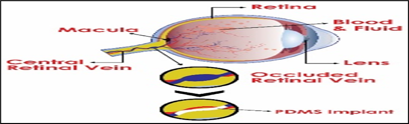
Figure 5. Proposed Microchannel Implantation to Treat RVO
Artificial intelligence software enabled the researcher to deal with complex problems uncomplicatedly. Contrary to binary logic fuzzy logic provided a more accurate picture of the system by considering all possibilities. Instead of overlooking at the implications of minutia, the fuzzy technique comprehensively enveloped all possible happenings. Fuzzy logic was beneficial in managerial sciences, business research, economics, scientific research, and various other engineering and social sciences fields.
In MEMS technology mainly micro-size components are dealt with, where simulations of possible happening allow to save time, money, and energy for the process. Researchers have applied fuzzy techniques in the field of microfluidics to manage the fabrication and operation of devices [42]. The fuzzy simulation depicted fluid flow properties and related outcomes well in advance. Morphology and geometry of micro-channels and other research employed in microfluidic devices were being predicted by fuzzy and ANSYS simulation for perfect outcomes [20, 43–49].
In the current study, four types of micro-channels architecture sinusoidal, U-shaped, spiral, and curvilinear were subjected to fuzzy parametric estimations to analyze the suitability for biomedical implantation to treat RVO. This microchannel architecture was selected to see the morphology of retinal veins. Moreover, the material used for fabrication can augment the evaluation of perfect-fit choices.
For this purpose, four Fuzzy Logic Control (FLC) models one for each microchannel was prepared. Membership functions for each model was defined and rules were prepared by using “if and then” statements.
The outcomes are discussed below for all three sinusoidal, curvilinear, and U-shaped microchannel.
For sinusoidal microchannel, Fuzzy Inference System (FIS) variables and their ranges are shown in Table 1.
Table 1. FIS Variables and their Ranges for Sinusoidal Microchannel
|
Input FIS Variables and Their Ranges |
Output FIS Variables with Their Ranges |
|||
|
Pressure |
Diameter |
Reynold Number |
Flow Rate |
Velocity |
|
10-100 KPa |
100-1000 µm |
100-200 |
1-3 nL/s |
40-60 mm/s |
The output critical for a retinal vein is flow rate and velocity. The range of Reynolds numbers was taken to take all possibilities into account.
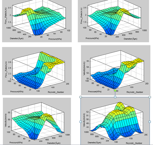
Figure 6. 3D Representation of Variations of Velocity and Flow rate in Sinusoidal microchannel
Flow rate showed a great variance in response to varying pressure. Only for pressure near 85 KPa flow rate remained in the acceptable value near 2nL/s. Response of flow rate to variations in Reynold's number and diameter of the microchannel was spread over a wide range, showing variation in inputs for each unit change. This variation in the flow rate was a response to minor changes in all three input parameters, which made this the least compatible for the intended purpose. Velocity variations follow the same pattern of changes, which were followed in response for the input changes in the microchannel simulation.
Fore spiral microchannel Fuzzy Inference System (FIS) variables and their ranges are shown in Table 2.
Table 2. FIS Variables and their Ranges for Spiral Micro-channels
|
Input FIS Variables and Their Ranges |
Output FIS Variables with Their Ranges |
|||
|
Pressure |
Diameter |
Friction |
Flow Rate |
Velocity |
|
20-100 KPa |
100-800 µm |
0-1 N |
1-3 nL/s |
40-60 mm/s |
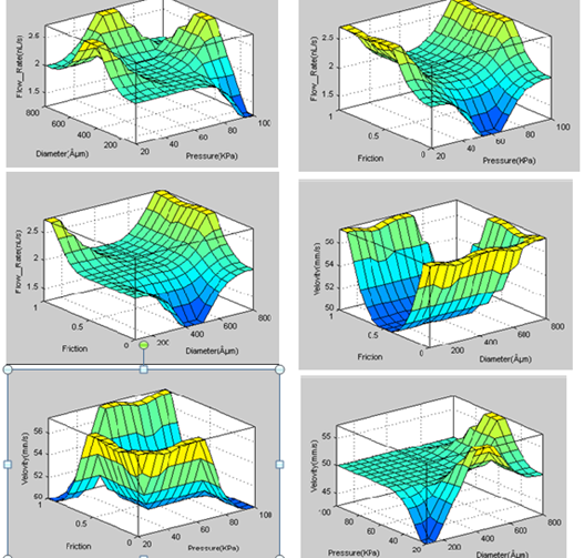
Figure 7. 3D Representation of Variations of Velocity and Flow rate in Spiral Microchannel
For variations in pressure, the flow rate varies to 2-2.75 nL/S. Only for 40-80 KPa pressure ranges, the flow rate remained in acceptable ranges near 2.1 nL/s. whereas in larger diameters the flow rate tends to increase constantly. Moreover, flow rate and velocity showed the same response of variations in the diameter of the microchannel. However, velocity remained constant near 5 mm/s for the pressure range of 55-10 KPa.
Fore U-Shaped microchannel Fuzzy Inference System (FIS) variables and their ranges are shown in Table 3.
Table 3. FIS Variables and their Ranges for U-Shaped Micro-Channel
|
Input FIS Variables and Their Ranges |
Output FIS Variables with Their Ranges |
||
|
Pressure |
Diameter |
Velocity |
Flow Rate |
|
10-100 KPa |
100-1000 µm |
1-3 nL/s |
40-60 mm/s |
For low diameters up to 600 µm, the flow rate maintains a constant value of 2nL/s, however, an increased flow rate was noticed in the range of 600-950 µm that goes up to 2.7 nL/s. Contrarily, all pressures flow rate remained constant near 2nL/s. Minor changes in pressure and diameter made velocity vary to a great extent.

Figure 8. 3D Representation of Variations of Velocity and Flow rate in U-Shaped Microchannel
Fore U-Shaped microchannel Fuzzy Inference System (FIS) variables and their ranges are shown in Table 4.
Table 4. FIS Variables and their Ranges
|
Input FIS Variables and Their Ranges |
Output FIS Variables and Their Ranges |
|||
|
Pressure |
Diameter |
Friction |
Flow Rate |
Velocity |
|
20-100 KPa |
100-800 µm |
0-1 N |
1-3 nL/s |
40-60 mm/s |
Curvilinear micro-channels possess the quality of U-Shape and sinusoidal micro-channels. Moreover, its morphological appearance resembles the natural retinal veins, which gives it a clear advantage over the other types.
From the graphical representation it is quite obvious that for a diameter in the range of 400-600µm its flow rate and velocity exhibit the ideal optimization. The most suitable situation in the curvilinear microchannel is that in the pressure range 50-100 KPa flow rate remains constant near 2 nL/s. Moreover, the flow rate also maintains a constant value near 2nL/s when the diameter of the channel ranges between 400-100 µm. Velocity for any certain diameter maintains a constant value, which is ideal for the proposed application.
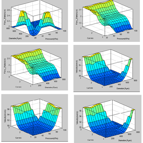
Figure 9. 3D Representation of Variations of Velocity and Flow rate in Curvilinear Microchanne
3.5. Comparative Discussion
A comparison of most optimum values was figured out through fuzzy parametric estimations, which is given below in Table 5. Both the flow rate and the velocity were compared with the Mamdani values to find the percentage errors. Fuzzy rule views provided us with the ease to see all possible parameters for the optimal outputs.
Although the percentage error in all four cases was quite negligible, the actual blood velocity and average flow rate values 54 mm/s and 2.04 nanoliters/ sec, respectively fall close in all simulated microchannels.
Table 5. Flow Rate and Velocity Results
|
Microchannel Type |
Quantity |
FUZZY Value |
Mamdani Value |
%age Error |
|
Sinusoidal |
Flow Rate |
2.18 nL/s |
2.15 nL/s |
1.4% |
|
Velocity |
50.7 mm/s |
50.5 mm/s |
0.4% |
|
|
Spiral |
Flow Rate |
2.19 nL/s |
2.16 nL/s |
1.4% |
|
Velocity |
51.6mm/s |
51.9mm/s |
0.6% |
|
|
U-Shape |
Flow Rate |
2.0 nL/s |
2.3 nL/s |
1.5% |
|
Velocity |
50 mm/s |
50.2 mm/s |
0.4% |
|
|
Curvilinear |
Flow Rate |
2.06 nL/s |
2.04 nL/s |
0.1% |
|
Velocity |
51.8 mm/s |
51.5 mm/s |
0.6% |
The current study aimed to provide a fluidic stimulation by using Fuzzy parametric estimations technique, which provided optimal behaviours for all four micro-channels. The output optimization of four simulated micro-channels revealed that the curvilinear micro-channel best suited the implantation to treat RVO. Blood flow rate and velocity are the least divergent in a curvilinear microchannel. Although the simulated values of curvilinear micro-channels are a little lower than the practical value, this small deficiency is negligible in the context of associated flow rate. For the three simulations, the combination of two output parameters remained divergent from the actual case requirements.