
| Review | Open Access |
|---|
Polymeric Rod-shaped Lanthanum Oxide Nanoparticles Generated from Polygonum Minus Leaf Extract: Synthesis, Characterization, and Antioxidant Activities |
 |
|---|
1. INTRODUCTION
In recent decades, green synthesis of metal oxide nanoparticles has been studied broadly due to their high surface to volume ratio. Among the other metal oxide nanoparticles, the oxides of rare-earth elements show various fascinating characteristics, including a high number of active sites, significant structural stability, as well as superior optical, magnetic, and chemical capabilities [1]. The lanthanoids-based nanoparticles garner a lot of attention due to their wide range of uses in biomedicine, chemical industry, and agriculture and material sciences [2]. In addition, lanthanoids can also be applied in X-ray tomography imaging, near infrared (NIR) imaging, single-photon emission computed tomography (SPECT), biosensing, and antioxidant therapy [3].
Numerous methodologies and protocols are available for the synthesis of La2O3 NPs, including solvothermal, hydrothermal, chemical precipitation, and green synthesis. Among these, green synthesis provides environmentally friendly products by removing or reducing the harmful substances [4]. Green nanotechnology scholars have studied the synthesis of nanoparticles thoroughly using various microorganisms, such as algae, fungi, bacteria, and viruses [5]. Moreover, metal oxide nanoparticles produced using plant extracts have useful practical applications [6]. The several silver oxide NPs produced using tridax procumbens and cluster bean extracts are used as biosensors, revealing their high antimicrobial activity [7]. Furthermore, platinum oxide NPs obtained from the holy basil extract are used for catalytic activity [8]. Moreover, lanthanum nanoparticles produced using multingia calabura leaves extract are used for antibacterial activity against Staphylococcus aureus and Escherichia coli. These nanoparticles exhibit good anticoagulant, thrombolytic, and hemolytic activities with antioxidant inhibition of 70.06% [6]. In addition, lanthanum oxide nanoparticles (La₂O₃ NPs) produced by Andrographis paniculata leaf extract show promising antibacterial activity against S. aureus and E. coli, as well as anti-inflammatory and anticancerous properties [9]. La₂O₃ NPs produced using Eucalyptus globulus leaf extracts are very useful to control various inflammations and diabetic diseases [10]. Similarly, green synthesis of La₂O₃ NPs with the help of different plant extracts (Physalis angulate, Datura metel, Muntingia calabura, Andrographis paniculate, Vigna radiata, Trigonella and foenum-graecum) has remarkable applications in electronics, biomedicine, insulators, and biocatalysts [11].
Based on the aforementioned studies, plant-mediated synthesis of La₂O₃ NPs has received very little consideration by scientific community, although there is a broad scope for La₂O₃ NPs using various plant extracts. Polygonum minus extract has been used for the synthesis of noble metal nanoparticles (e.g., Au, Ag), although its application to rare-earth compounds, such as La₂O₃ NPs, remains novel. In this regard, the preparation of La₂ O₃ NPs is a groundbreaking step toward making rare-earth nanoparticles in an environmentally friendly way. This method makes use of the extract’s flavonoid, phenolic acid, and terpene content as dual-purpose agents [12], allowing for the near-complete reduction of La2+ ions in 10 minutes at room temperature. This improves the efficiency of the synthesis by removing the need for energy-intensive procedures, such as high pressure or temperature. Additionally, by avoiding dangerous chemicals like NaBH₄ or organic solvents, reducing waste production, and producing biocompatible, low-cytotoxic NPs appropriate for biomedical applications—as demonstrated by smaller ecological footprints in similar green syntheses—it also supports environmental sustainability [13]. This approach not only lowers pollution and energy use but also conforms to eco-friendly nanotechnology principles, creating opportunities for the economical, scalable manufacture of rare-earth NPs with a negligible environmental impact. Hence, the current study focuses on the synthesis of La₂O₃ NPs using P. minus leaf extract. The synthesized nanoparticles are further investigated for their antioxidant activities.
2. METHODS
2.1. MaterialsAll the chemicals employed in the research were of analytical grade and purchased through suppliers. Lanthanum (III) nitrate hexahydrate (La(NO3)3.6H2O) was obtained from Sigma-Aldrich. Whereas, fresh leaves of P. minus were purchased from a local market of Malaysia.
2.2. InstrumentationThe synthesized La2O3 NPs were characterized using various analytical tecniques including Uv- Vis spectroscopy, in the range of 400-800 nm, using Ultra-3000 series spectrophotometer. The amorphous structure of La2O3 NPs was determined by XRD (D2 Phaser from BRUKER Germany model A26-X1-A2B0B2A0). While, the FTIR analysis of La2O3 NPs was carried out via Perkin Elmer Spectrometer 1600, in the region of 400-4000 cm-1, by using standard KBr pellet technique. The presence of elemental lanthanum, as well as its morphology, was determined by using EDX coupled with FESEM (Thermo Scientific model Apreo 2s). The average particle size was determined with the help of image j software. Thermal activity (TGA) was determined in a nitrogen atmosphere with a heating rate of 10oC/min using TA instruments Q50 TGA analyzer.
2.3. Preparation of Plant Extract SolutionA total of 1.0 g of P. minus leaf powder dried in an oven at 35oC was boiled in 50 ml of de-ionized water for 15 minutes before filtration. The filtered extract was refrigerated at 4°C. This extract was employed as a reducing and stabilizing agent in the preparation of nanoparticles.
2.4. Preparation of Lanthanum Salt SolutionApproximately 1mm solution of lanthanum (III) nitrate hexahydrate (La (NO3)3.6H2O) was prepared by adding 0.01 g of lanthanum salt in 30 ml of de-ionized water.
2.5. Synthesis of Lanthanum Oxide NanoparticlesA total of 9 ml leaf extract solution was added dropwise into the salt solution (30 ml; 1 mm). The mixture was stirred for 20 mins. The resulting solution was centrifuged at 16000 rpm for another 20 mins. The resultant pellets were re-dispersed into de-ionized water and methanol after the supernatants were discarded. To get rid of any impurities adsorbed on the surface of La₂O₃ NPs, the centrifugation procedure was repeated two to three times and the powder was dried in a hot air oven at 45ᴼC.
2.6. Antioxidant Activity>Antioxidants are chemicals, either natural or artificial, that stop or postpone cell damage brought on by oxidants. Any chemical that delays, stops, or eliminates oxidative damage in the target molecule is an antioxidant. The chemical must be active at low radical concentrations in order to be regarded as an antioxidant. Its amount must be sufficient to deactivate the target molecule. Further, it must react with oxygen or nitrogen free radicals and the final product of the reaction must be less toxic than the removed radical. Phenolic antioxidants frequently lose their activity at high concentrations and act as prooxidants. Various antioxidants interact with diverse reactive species in a variety of ways at different sites, while defending certain biological targets [13, 14].
2.6.1. Preparation of Samples for Antioxidant Activity. For antioxidant activity, three different types of stock solutions were prepared including La2O3 NPs and DPPH solutions, as well as ascorbic acid solution. The free radical scavenging activity of La2O3 NPs and conventional ascorbic acid was tested using the stable radical DPPH (0.004g/100ml methanol) solution. Approximately 5mg/ml of La2O3 NPs and a similar amount of ascorbic acid at various concentrations (10, 20, 30, 50, 100, and 200 mg/ml) were vortexed violently with 3 ml freshly produced DPPH solution. The solution was then incubated at room temperature in the dark for 30 minutes. A Uv-Vis spectrophotometer at 517 nm was used to measure absorption. DPPH was used as control, while methanol was used as blank solution [14].
Scavenging% = Hc-Hs/Hc 100
Here, Hc is the absorbance of control (DPPH) and Hs is the absorbance of La2O3 NPs / ascorbic acid.
3. RESULTS AND DISCUSSION
3.1. Uv-Visible AnalysisThe size, shape, and morphology of La2O3 NPs are greatly influenced by a number of factors, including pH, temperature, reaction time, and concentration. Their synthesis and stability between 200-800 nm were evaluated using a Shimadzu spectrophotometer [15].
3.1.1 Time Dependent Studies. Figure 1 displays the absorption spectra of La₂O₃ NPs synthesized using 1 mm lanthanum salt solution and 9 ml P. minus leaf extract, measured at various time intervals. A stable absorption peak at 300-400 nm indicates successful NP synthesis, with no significant change in intensity over two weeks, confirming the long-term stability of the La₂O₃ NPs [16].
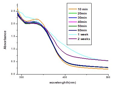
Figure 1. UV-Vis Absorption Spectra of La₂O₃ NPs Over Time
3.1.2. Concentration Dependent Studies. The effects of the concentrations of leaf extract and salt solution [17] are depicted in Figures 2a and 2b, respectively. Figure 2a shows the effects of 9, 18, and 27 ml leaf extract concentrations on (1 mm) salt solution. As the amount of leaf extract increased, a gradual decrease was observed in the peak of La2O3 NPs, as evident by the Uv-Vis spectra of the resulting particles. It was found that adding 9 ml of leaf extract to the reaction mixture was successful in producing La2O3 NPs. Uv-Vis spectra showed that the sharpness of the absorption peak is dependent on the leaf extract concentration, thus the peak was sharper at 9 ml. However, with the increase in leaf extract red shift was recorded, indicating an increase in particle size [16].
Furthermore, Figure 2b shows the effects of 1, 2, and 3 mm salt solutions respectively on 9 ml of leaf extract in the synthesis of La2O3 NPs. It was noticed that all the selected concentrations were effective in the synthesis of La2O3 NPs and a clear band was observed at 308 nm [17]. Hence, it was proved that the concentration of leaf extract can affect synthesis La2O3 NPs production, as compared to the concentration of salt solution.
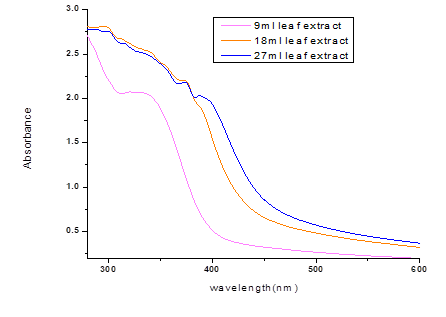
Figure 2a. UV-Vis Spectra Showing the Effects of Leaf Extract Concentration on La₂O₃ NP Synthesis
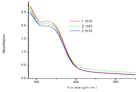
Figure 2b. UV-Vis Spectra Showing the Effects of Lanthanum Salt Concentration on La₂O₃ NP Synthesis
3.1.3. Temperature Dependent Studies. The size and morphology of La2O3 NPs were greatly affected by the reaction temperature. Figure 3 depicts the absorption spectra of La2O3 NPs produced at varied temperatures, ranging from room temperature to 80°C. Notably, the absorption peak and wavelength did not change with an increase in temperature. It was fascinating to see that synthesized La2O3 NPs remained extremely stable, even at a high temperature [16].
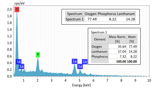
Figure 3. UV-Vis Spectra of La₂O₃ NPs at Different Temperatures (RT to 80°C)
3.1.4. pH Dependent Studies. The pH is another important factor which affects the size, shape, and morphology of the synthesized La2O3 NPs. The electrical charges of biomolecules can alter as pH changes, which may have an impact on their reducing ability, capping ability, and growth [18]. The impact of both acidic and basic pH on the absorption spectra of the synthesized La2O3 NPs is depicted in Figures 4a and 4b. Figure 4a reveals that when the pH dropped, a clear band of nanoparticles was seen even at the lowest pH. Whereas, Figure 4b shows that a rise in pH caused the absorption peak to shift towards a longer wavelength (from 308 to 450 nm), indicating that the size of the La2O3 NPs produced increased while their production decreased, suggesting the existence of larger particles with a polydispersed distribution. This may partially be due to the reaction of sodium hydroxide and the impact of acidic phenolic groups on flavonoids in the extract. Such an increase in particle size, coupled with an increase in the pH value, has been reported previously [19–21]. Therefore, the production of La2O3 NPs is more favored by an acidic pH as compared to a basic pH.
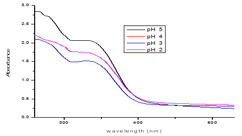
Figure 4a. UV-Vis Spectra of La₂O₃ NPs at Various Acidic pH Levels
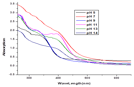
Figure 4b.UV-Vis Spectra of La₂O₃ NPs at Various Basic pH Levels
3.2. EDX AnalysisThe purity and weight percentage of the produced nanoparticles was confirmed by using the energy dispersive X-ray spectrometer. Figure 5 shows a representative EDX spectrum. Several peaks of La can be distinguished in addition to the signals for O and P. The observed O and La peaks are related to La2O3 NPs. The presence of P was extract-derived and no toxic elements were detected, as reported earlier [22, 23].
Figure 5. EDX Spectrum of La₂O₃ NPs
3.3. Powder X-ray Diffraction AnalysisP-XRD is used to determine amorphous nature and phase purity. The virtual broadband in the XRD pattern reveals the lack of periodic crystal structure in the amorphous sample, implying that the produced sample is entirely amorphous, as shown in Figure 6. It can be deduced that the synthesized La2O3 NPs exhibited a typical amorphous phase, as reported in the literature, while the crystallite size was not applicable for this study due to the lack of sharp crystalline peaks [24, 25]. This amorphous character, also observed in other biosynthesized metal oxide nanoparticles [25], may contribute to their enhanced surface reactivity. This is advantageous for antioxidant activity, as amorphous structures often provide more active sites for radical scavenging, as compared to their crystalline counterparts [6]. These results underscore the potential of P. minus-mediated La₂O₃ NPs for biomedical applications, particularly in antioxidant therapies.
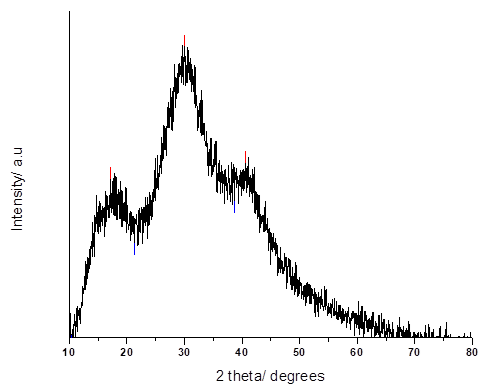
Figure 6. Powder XRD Pattern of La₂O₃ NPs
3.4. FE-SEM Studies"The size and surface morphology of La2O3 NPs were observed using FE-SEM. The images of La2O3 NPs are given in Figure 7a. It is clear that these nanoparticles are elongated and rod-like in their shape. They have a uniform particle size of about 343 nm, determined by image J software and shown in histogram (Figure 7b), confirming a rod-like morphology and polydispersity. Polymeric nanoparticles are particles measuring between 1-1000 nm in size. They showed a significant potential for targeted drug delivery during the treatment of various medical conditions. Moreover, they can also be used for a variety of antibacterial, antifungal, and cytotoxic purposes [26-29].

Figure 7a. FE-SEM Images of La₂O₃ NPs
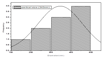
Figure 7b. Particle Size Distribution Histogram of La₂O₃ NPs
3.5. FTIR AnalysisThe FTIR analysis was carried out to identify the possible functional groups of biomolecules present in the P. minus leaf. These biomolecules are responsible for the reduction of La (NO3)3.6H2O to elemental lanthanum and stabilization of the formed La2O3 NPs. FTIR spectra of the P. minus leaf powder and the synthesized La2O3NPs, using 9 ml of P. minus extract at pH 5, is shown below in Figure 8a. The FTIR spectrum of leaf powder showed characteristic bands for O-H stretching vibrations at 3428 cm-1 (polyols), stretching vibrations of C=O at 1638cm-1 (unsaturated carbonyl group), and stretching vibrations of C-O at 1068 cm-1 (polyols) [30, 31]. The asymmetric stretching vibrations of C-H at 2924 cm-1, as well as the stretching vibrations of CHO at 1625 cm-1, confirmed the presence of phenolic compounds including flavonoids (quercetin and myricetin) in the P. minus leaf aqueous extract, as reported earlier [32].
Absorption sharp band clearly observed at 614 cm–1 is attributable to La-O stretching vibration, as depicted in Figure 8b, confirming the successful synthesis of La2O3 NPs [33, 34]. This observation suggests the likely involvement of the flavonoids of the P. minus leaf extract in the bio reduction process of La +3 to La (0), stabilizing the La2O3 NPs [35]. The presence of these bioactive molecules on the surface of nanoparticles likely enhanced their biocompatibility and contributed to their moderate antioxidant activity (28% DPPH scavenging at 200 μg/ml), since phenolic compounds are known to donate electrons to neutralize free radicals, a mechanism supported by research on plant-mediated nanoparticle synthesis [17].
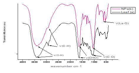
Figure 8a. FTIR Spectra of P. minus Leaf Powder and Synthesized La2O3 NPs
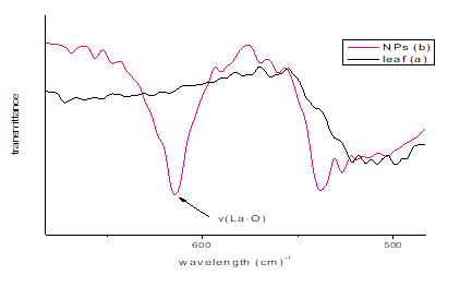
Figure 8b. FTIR Spectra Showing La-O Band in La₂O₃ NPs
3.6. Thermal Gravimetric AnalysisThermal studies were performed to investigate lanthanum nanoparticles, where thermogravimetric analysis was executed within the temperature range of ambient to 900ᴼC. Thermal stability is regarded as a remarkable property of La2O3 NPs. Thermal curves of the compounds are given below in Figure 9. La2O3 NPs decomposed thermally in 3 main steps. Firstly, the weight loss of 10% in the temperature range 70-191ᴼC, with the DTA temperature of 75ᴼC, is attributable to the loss of crystal water. Secondly, a further decay through the combustion of organic moieties with a weight loss of 53% was observed at the temperature range of 191-380ᴼC, with the DTA temperature of 388ᴼC. Finally, decomposition occurred with the residual mass of 46% at the DTA temperature of 815ᴼC. This thermal stability, as reported in similar studies [33], indicates that the organic capping agents from P. minus enhance the nanoparticles’ structural integrity, which is crucial for their application in biological environments where thermal and chemical stability are required.
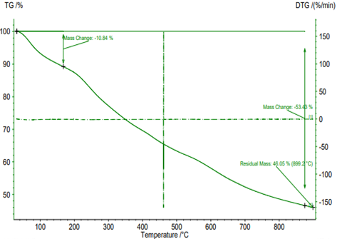
Figure 9. TGA/DTA Curves of La₂O₃ NPs
3.7. Antioxidant ActivitiesDPPH radical scavenging assay 2,2-diphenyl-1-picrylhydrazyl is a stable free synthetic radical at room temperature. Further, by accepting an electron or hydrogen radical, it becomes a stable molecule. In the DPPH assay, antioxidants reduced the DPPH radical to the non-radical form. Hence, absorption was reduced and the color of the DPPH solution changed from purple to yellow. This is known as scavenging and it can be done only by an antioxidant. In the current study, when the sample was subjected to DPPH, 28% of scavenging occurred. The DPPH scavenging of the La2O3 NPs confirmed its antioxidant nature. Lanthanum nanoparticles showed antioxidant activity but less than standard ascorbic acid, as observed in Figures 10a and b.
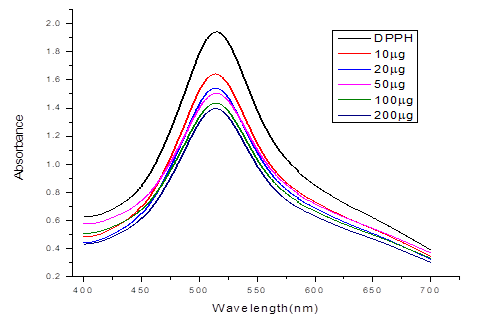
Figure 10a. DPPH Scavenging Activity of La₂O₃ NPs
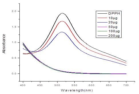
Figure 10b. DPPH Scavenging Activity of Ascorbic Acid
The percentage of free radical scavenging activity of various samples at different concentrations ranging from 10 to 200 µg/ml for standard ascorbic acid is shown below in Figure 11 [36].
La₂O₃ NPs, synthesized by P. minus, exhibited a DPPH scavenging activity of 28% at 200 μg/ml, demonstrating moderate antioxidant potential. This performance is comparably moderate as compared to other nanoparticles synthesized by different plant extracts, as reported in the literature. For instance, among plant-mediated lanthanum NPs, those synthesized using Muntingia calabura-derived lanthanum NPs showed a scavenging activity of 70.06% at 5 mg/ml. Similarly, silver nanoparticles synthesized from Petiveria alliacea L. leaf extract displayed antioxidant activity ranging from 61.39% to 70.69% at 5 mg/ml [6].
Scavenging activity depends on the quality and quantity of the antioxidant (nanoparticles). As stated above, La₂O₃ NPs showed moderate quality as compared to other nanoparticles mentioned above. The antioxidant activity of lanthanum nanoparticles may be attributed to bioactive chemicals, primarily polyphenols, that are coated on them. According to early reports, lanthanum nanoparticles made from plant extracts have antioxidant properties and their antioxidant nature grows as their concentration rises. Hence, if the concentration of La₂O₃ NPs increases, its antioxidant activity may gradually increase as well [6].
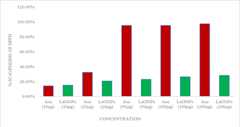
Figure 11. DPPH Scavenging Percentages for La₂O₃ NPs and Ascorbic Acid
The study revealed that at the concentration of 200μg/ml, La₂O₃ NPs showed greater antioxidant activity by scavenging 28% DPPH but less than ascorbic acid, that is, 97%. Percentage scavenging of DPPH revealed the potency of the sample towards its antioxidant activity.
Table 1 shows the DPPH scavenging ability of lanthanum nanoparticles and ascorbic acid at varied concentrations.
Table 1. DPPH Scavenging Percentages at Various Concentrations
|
Samples |
Methanol |
DPPH |
Absorbance |
% Scavenging of DPPH |
|---|---|---|---|---|
|
Ascorbic acid (10 μg/ml) |
990 μl |
3 ml |
1.66 |
14.3% |
|
Ascorbic acid (20 μg/ml) |
980 μl |
3 ml |
1.31 |
32.3% |
|
Ascorbic acid (50 μg/ml) |
950 μl |
3 ml |
0.09 |
95.3% |
|
Ascorbic acid (100 μg/ml) |
900 μl |
3 ml |
0.06 |
95.2% |
|
Ascorbic acid (200 μg/ml) |
800 μl |
3 ml |
0.05 |
97.4% |
|
NPs (10 μg/ml) |
990 μl |
3 ml |
1.644 |
15% |
|
NPs (20 μg/ml) |
980 μl |
3 ml |
1.537 |
20.6% |
|
NPs (50 μg/ml) |
950 μl |
3 ml |
1.494 |
22.8% |
|
NPs (100 μg/ml) |
900 μl |
3 ml |
1.422 |
26.5% |
|
NPs (200 μg/ml) |
800 μl |
3 ml |
1.388 |
28.3% |
This research established a simple and easy technique of synthesizing polymeric La2O3 NPs from the aqueous extract of P. minus. The effect of temperature, reaction time, concentration, and pH on La2O3 NPs were analyzed by Uv-Vis spectroscopy. A clear band of La-O nanoparticles at 308 nm was observed. The peaks in the FTIR spectra indicated the potential role of different functional groups of plant metabolites as capping and stabilizing agents, while nanoparticles showed the characteristic band of La-O at 614 cm-1. The size of the synthesized nanoparticles was 343 nm, as determined by image J software. EDAX analysis confirmed the presence of lanthanum along with oxides and phosphorus. While, XRD pattern confirmed the amorphous nature of lanthanum nanoparticles. Further, TG-DTA analysis was performed to investigate the thermal properties of La2O3 NPs. The synthesized La2O3 NPs were found to exhibit moderate antioxidant activity. It can be concluded that further in vitro and in vivo explorations can fully assess the biomedical applicability of these synthesized polymeric nanoparticles, particularly in areas such as imaging, sensing, therapy, and antimicrobial activities, as demonstrated in biocompatibility assessments and potential applications of lanthanide nanomaterials.
3.9. Future DirectionFuture research should concentrate on thorough in vitro studies to assess the biocompatibility, cytotoxicity, and potential biomedical applications of polymeric lanthanum oxide nanoparticles (La2O3 NPs) synthesized using P. minus leaf extract, including targeted drug delivery and antimicrobial activity. Investigating different plant extracts with rich phytochemical profiles, such as Ocimum sanctum or Azadirachta indica, may improve the manufacturing process, as well as the stability and bioactivity of nanoparticles.
CONFLICT OF INTEREST
The authors declare that they have no known competing financial interests or personal relationships that could have appeared to influence the work reported in this paper.
DATA AVAILABILITY STATEMENT
All data generated and analyzed during this study are included in this article.
FUNDING DETAILS
No funding has been received for this research.
REFERENCES
- Sulaiman N, Yulizar Y, Apriandanu DOB. Eco-friendly method for synthesis of La2O3 nanoparticles using Physalis angulata leaf extract. AIP Conf Proc. 2018;(1):e020105. https://doi.org/10.1063/1.5064102
- Hollande E, Lebugle A. Europium-doped bioapatite: a new photostable biological probe, internalizable by human cells. Biomater. 2003;24:3365–3371. https://doi.org/10.1016/S0142-9612(03)00169-8
- Balusamy B, Kandhasamy YG, Senthamizhan A, Chandrasekaran G, Subramanian MS, Kumaravel TS. Characterization and bacterial toxicity of lanthanum oxide bulk and nanoparticles. J Rare Earths. 2012;30(12):1298–1302. https://doi.org/10.1016/S1002-0721(12)60224-5
- Moothedan M, Sherly KB. Synthesis, characterization and sorption studies of nano lanthanum oxide. J Water Process Eng. 2016;9:29–37. https://doi.org/10.1016/j.jwpe.2015.11.002
- Akhtar MS, Panwar J, Yun YS. Biogenic synthesis of metallic nanoparticles by plant extracts. ACS Sustain Chem Eng. 2013;1(6):591–602. https://doi.org/10.1021/sc300118u
- Kumar KM, Hemananthan E, Devi PR, Kumar SV, Hariharan RJ. Biogenic synthesis, characterization and biological activity of lanthanum nanoparticles. Mater Today Proc. 2020;21:887–895. https://doi.org/10.1016/j.matpr.2019.07.727
- Narayanan KB, Sakthivel N. Green synthesis of biogenic metal nanoparticles by terrestrial and aquatic phototrophic and heterotrophic eukaryotes and biocompatible agents. Adv Colloid Interface Sci. 2011;169(2):59–79. https://doi.org/10.1016/j.cis.2011.08.004
- Zheng H, Zhu K, Onda A, Yanagisawa K. Hydrothermal synthesis of various shape-controlled europium hydroxides. Nanomater. 2021;11(2):1–10. https://doi.org/10.3390/nano11020529
- Veerasingam M, Murugesan B, Mahalingam S. Ionic liquid mediated morphologically improved lanthanum oxide nanoparticles by Andrographis paniculata leaves extract and its biomedical applications. J Rare Earths. 2020;38(3):281–291. https://doi.org/10.1016/j.jre.2019.06.006
- Maheshwaran G, Muneeswari RS, Bharathi AN, Kumar MK, Sudhahar S. Eco-friendly synthesis of lanthanum oxide nanoparticles by Eucalyptus globulus leaf extracts for effective biomedical applications. Mater Lett. 2021;283(1):e128799. https://doi.org/10.1016/j.matlet.2020.128799
- Dabhane H. Plant mediated green synthesis of lanthanum oxide (La2O3) nanoparticles: a review. Asian J Nanosci Mater. 2020;3:291–299. https://doi.org/10.26655/AJNANOMAT.2020.4.3
- Miean KH, Mohamed S. Flavonoid (myricetin, quercetin, kaempferol, luteolin, and apigenin) content of edible tropical plants. J Agric Food Chem. 2001;49(6):3106–3112. https://doi.org/10.1021/jf000892m
- Ingale AG, Chaudhari AN. Biogenic synthesis of nanoparticles and potential applications: an ecofriendly approach. J Nanomed Nanotechnol. 2013;4(2):e1000165. https://doi.org/10.4172/2157-7439.1000165
- Shameli K, Bin Ahmad M, Al-Mulla EAJ, et al. Green biosynthesis of silver nanoparticles using Callicarpa maingayi stem bark extraction. Molecules. 2012;17(7):8506–8517. https://doi.org/10.3390/molecules17078506
- Bhakya S, Muthukrishnan S, Sukumaran M, Muthukumar M. Biogenic synthesis of silver nanoparticles and their antioxidant and antibacterial activity. Appl Nanosci. 2016;6(5):755–766. https://doi.org/10.1007/s13204-015-0473-z
- Saware K, Venkataraman A. Biosynthesis and characterization of stable silver nanoparticles using Ficus religiosa leaf extract: a mechanism perspective. J Clust Sci. 2014;25(4):1157–1171. https://doi.org/10.1007/s10876-014-0697-1
- Jain S, Mehata MS. Medicinal plant leaf extract and pure flavonoid mediated green synthesis of silver nanoparticles and their enhanced antibacterial property. Sci Rep. 2017;7:e15867. https://doi.org/10.1038/s41598-017-15724-8
- Dwivedi AD, Gopal K. Biosynthesis of silver and gold nanoparticles using Chenopodium album leaf extract. Colloids Surf Physicochem Eng Asp. 2010;369(1–3):27–33. https://doi.org/10.1016/j.colsurfa.2010.07.020
- Liu B, Xie J, Lee JY, Ting YP, Chen JP. Optimization of high-yield biological synthesis of single-crystalline gold nanoplates. J Phys Chem B. 2005;109(32):15256–15263. https://doi.org/10.1021/jp051449n
- Panáček A, Kvitek L, Prucek R, et al. Silver colloid nanoparticles: synthesis, characterization, and their antibacterial activity. J Phys Chem B. 2006;110(33):16248–16253. https://doi.org/10.1021/jp063826h
- Noruzi M, Zare D, Davoodi D. A rapid biosynthesis route for the preparation of gold nanoparticles by aqueous extract of cypress leaves at room temperature. Spectrochim Acta A Mol Biomol Spectrosc. 2012:84–88. https://doi.org/10.1016/j.saa.2012.03.041
- Diallo A, Beye AC, Doyle TB, Park E, Maaza M. Green synthesis of Co3O4 nanoparticles via Aspalathus linearis: physical properties. Green Chem Lett Rev. 2015;8(3–4):30–36. https://doi.org/10.1080/17518253.2015.1082646
- Thema FT, Manikandan E, Dhlamini MS, Maaza MJ. Green synthesis of ZnO nanoparticles via Agathosma betulina natural extract. Mater Lett. 2015;161:124–127. https://doi.org/10.1016/j.matlet.2015.08.052
- Xing J, Yan F, Zhao Y, et al. High-efficiency light-emitting diodes of organometal halide perovskite amorphous nanoparticles. ACS Nano. 2016;10(7):6623–6630. https://doi.org/10.1021/acsnano.6b01540
- Hoang V, Ganguli D. Amorphous nanoparticles - experiments and computer simulations. Phys Rep. 2012;518(3):81–140. https://doi.org/10.1016/j.physrep.2012.07.004
- Khan I, Saeed K, Khan I. Nanoparticles: properties, applications and toxicities. Arab J Chem. 2019;12(7):908–931. https://doi.org/10.1016/j.arabjc.2017.05.011
- Lee H, Lytton-Jean AK, Chen Y, et al. Molecularly self-assembled nucleic acid nanoparticles for targeted in vivo siRNA delivery. Nat Nanotechnol. 2012;7(6):389–393. https://doi.org/10.1038/nnano.2012.73
- Carreiró F, Oliveira AM, Neves A, et al. Polymeric nanoparticles: production, characterization, toxicology and ecotoxicology. Molecules. 2020;25(16):e3731. https://doi.org/10.3390/molecules25163731
- Medina C, Santos‐Martinez MJ, Radomski A, Corrigan OI, Radomski MW. Nanoparticles: pharmacological and toxicological significance. Br J Pharmacol. 2007;150(5):552–558. https://doi.org/10.1038/sj.bjp.0707130
- Pocurull E, Marcé RM, Borrull F. Determination of phenolic compounds in natural waters by liquid chromatography with ultraviolet and electrochemical detection after on-line trace enrichment. J Chromatogr A. 1996;738(1):1–9. https://doi.org/10.1016/0021-9673(96)00070-2
- Dauthal P, Mukhopadhyay M. Prunus domestica fruit extract-mediated synthesis of gold nanoparticles and its catalytic activity for 4-nitrophenol reduction. Ind Eng Chem Res. 2012;51(40):13014–13020. https://doi.org/10.1021/ie300369g
- Miean KH, Mohamed S. Flavonoid (myricetin, quercetin, kaempferol, luteolin, and apigenin content of edible tropical plants. J Agric Food Chem. 2001;49:3106–3112.
- Imanaka N, Masui T, Kato Y. Preparation of the cubic-type La2O3 phase by thermal decomposition of LaI3. J Solid State Chem. 2005;178(1):395–398. https://doi.org/10.1016/j.jssc.2004.11.006
- Chakraborty P, Dam D, Abraham J. Bioactivity of lanthanum nanoparticle synthesized using Trigonella foenum-graecum seed extract. J Pharm Sci Res. 2016;8(11):1253–1257.
- Borhamdin S, Shamsuddin M, Alizadeh A. Biostabilised icosahedral gold nanoparticles: synthesis, cyclic voltammetric studies and catalytic activity towards 4-nitrophenol reduction. J Exp Nanosci. 2016;11(7):518–530. https://doi.org/10.1080/17458080.2015.1090021
- Singh J, Dhaliwal AS. Novel green synthesis and characterization of the antioxidant activity of silver nanoparticles prepared from Nepeta leucophylla root extract. Anal Lett. 2018;52(2):213–230. https://doi.org/10.1080/00032719.2018.1454936