

| Review | Open Access |
|---|
In silico Discovery of Cefoperazone as a Novel MMP-2 Inhibitor for Ovarian Cancer Therapy |
 |
|---|
In silico drug repurposing is a promising methodology to identify novel therapeutic applications for existing pharmaceuticals. This study explored the potential of eight third-generation cephalosporin antibiotics as matrix metalloproteinase-2 (MMP-2) allosteric inhibitors for selective targeting in ovarian cancer. MMP-2 is recognized for its pivotal role in tumor progression and metastasis, rendering it a compelling target for oncological treatment. We assessed the binding affinity and stability of selected cephalosporins with MMP-2 using molecular docking and molecular dynamics simulations. Our findings indicate that only Cefoperazone demonstrates robust binding interactions (∆G -8.1 Kcal/mol) within the active site of MMP-2, suggesting its viability as an effective inhibitor. Molecular dynamics (MD) simulations using normal mode analysis (NMA) further revealed that the Cefoperazone-MMP-2 complex is exceptionally stable under virtual physiological conditions. Complex stability was supported by a low B-factor and a minimal eigenvalue (1.51×10⁻⁴). Elastic network model analysis indicated proper molecular mobility close to the binding site, with atoms connected by flexible springs. Consequently, these findings offer novel insights into cefoperazone as a potential candidate for MMP-2 inhibition in the treatment of ovarian cancer. This research validates in silico methodologies for drug repurposing and supports the development of targeted therapeutic strategies for ovarian cancer.
1. INTRODUCTION
Ovarian cancer (OC) is the third most prevalent and deadly cancer of the female reproductive system [1]. Extracellular cell matrix (ECM) degradation, predominantly performed by Matrix Metalloproteinases (MMPs), namely, matrix metalloproteinase 2 (MMP-2), is one of the most popular causes of the invasiveness and metastasis of ovarian cancer [2].
MMP-2, a zinc-dependent endopeptidase, that degrades type IV and V collagen in the extracellular scaffold, is crucial in cancer progression [3]. Overexpression of MMP-2 in advanced-stage ovarian cancer is associated with adverse prognosis [4, 5], especially in stimulating angiogenesis and metastasis of malignancy cells. Therefore, MMP-2 inhibition acts as an attractive therapeutic option in ovarian cancer.
Computational techniques, such as drug repurposing and molecular docking, have dramatically transformed the roles of discovering inhibitors for MMP-2 as they provide considerable benefits compared with conventional drug discovery [6, 7]. These computational methods allow scientists to quickly scan through large databases of chemicals, significantly reducing the expenses and risks of conventional methodology and verifying the stage of drug development by pointing at early stages to preclinical and clinical trials [8, 9]. Molecular docking and virtual screening offer insights into interactions between the drug and target in detail, allowing the prediction of binding affinity, and optimizing baseline compounds before being tested [10]. In addition to speeding up the drug development process, this focused strategy raises the possibility of finding interesting candidates with higher efficacy and specificity.
It has been demonstrated that antibiotics prevent cancer development and induce apoptosis [11]. Various antibiotics have been investigated as potential treatment of cancer such as anthracyclines, mitomycin, bleomycin, actinomycin, and guanorycin [12]. Previous research has demonstrated the potential of cephalosporins in cancer therapy by sensitizing cells to cisplatin, activating the p53 pathway, and upregulating the expression of genes linked to apoptosis, such as HMOX1 and THBS1, in malignancies such as liver and nasopharyngeal carcinoma [13, 14].
Recent years have highlighted the potential use of antibiotics as MMP inhibitors within the scientific community. Varghese et al. [15] successfully employed these methods to investigate tetracycline compounds, such as eravacycline, as potential MMP inhibitors, proving the strength of in silico methods to study other new and repurposed drugs. Molecular docking and virtual screening studies also identified numerous possible ovarian cancer treatment agents by showing a promising level of binding to MMP-2 [16]. Therefore, through a combination of computational predictions and experimental interactions, investigators can find quicker ways to better chart out an increasingly complex territory of MMP-2 inhibition, which could reasonably give way to more practical therapeutic approaches to MMP-2-related diseases.
The present study contributes to the field of ovarian cancer treatment by exploring the repurposing potential of cephalosporins as possible allosteric inhibitors of matrix metalloproteinases (MMP-2), with noncatalytic regions targeted to enhance the specificity of the inhibitors. By systematically analyzing the binding affinities of eight cephalosporin molecules against MMP-2, this study not only identifies potentially effective drug candidates but also lays the foundation for new cost-efficient management approaches that can significantly transform the management of ovarian cancer and its outcome in patients.
1.MATERIALS AND METHODS
2.1.Protein Sequence RetrievalUniProt, a leading resource for protein sequences and functional annotation (https://www.uniprot.org/), was used to retrieve the MMP-2 protein FASTA sequence with UniProt ID P08253.
2.2.Physiochemical PropertiesTo determine the physiochemical properties of the selected protein, The ExPASy (Expert Protein Analysis) bioinformatics resource portal, established by the Swiss Institute of Bioinformatics. The ExPASy (Expert Protein Analysis) ProtParam tool was used to determine the physicochemical properties of the selected protein [17].
2.3. Secondary Structure PredictionPSIPRED database was used for the secondary structure prediction of the desired protein (http://bioinf.cs.ucl.ac.uk/psipred/). It is based upon information of the evolutionary related proteins that predict secondary structure of a novel amino acid sequence. A machine learning algorithm has been used to discover similar sequences and to create a position-specific scoring matrix that is then further refined by use of an artificial neural network that has been developed and trained to predict the secondary structure of the query sequence.
2.4. Tertiary Structure RetrievalPDB (Protein Data Bank) database was used to retrieve the tertiary structure of human MMP-2 (PDB ID: 1RTG) (https://www.rcsb.org/) which is hemopexin like regulatory domain lacking Zn+2 ion catalytic region. This database is home to the three-dimensional structural information of biological molecules like proteins and nucleic acids that have been deposited by biologists using experimental techniques like cryo-electron microscopy, NMR spectroscopy, and X-ray crystallography.
2.5. Refinement of the Protein ModelERRAT, a web-based tool, was used to assess the accuracy of protein structure derived from crystallography [18]. Further, Galaxy refine webserver (https://galaxy.seoklab.org/cgi-bin/submit.cgi?type=REFINE) was employed to achieve refined 3D structure of MMP-2 protein.
2.6. Verification of 3D StructureThe two-dimensional plot was generated through Ramachandran plot (https://swift.cmbi.umcn.nl/servers/html/ramaplot.html), a tool used to verify the protein structure. It displays the result in a chart form with torsion angles calculated of the protein with the values taken on the x-axis and the y-axis.
2.7Ligand RetrievalThe structure of eight cephalosporins used as inhibitors against the target MMP-2 was retrieved from the PubChem database (https://pubchem.ncbi.nlm.nih.gov/) (Table 1).
Table 1. Cephalosporins with PubChem IDs selected as Ligands
|
S no. |
Ligand Name |
th ID |
|---|---|---|
|
1 |
Cefixime |
5362065 |
|
2 |
Cefditoren |
9870843 |
|
3 |
Ceftibuten |
5282242 |
|
4 |
Cefoperazone |
44187 |
|
5 |
Ceftazidime |
5481173 |
|
6 |
Cefpodoxime |
6335986 |
|
7 |
Cefotaxime |
5742673 |
|
8 |
Ceftizoxime |
6533629 |
Molecular docking was performed using AutoDock Vina. The possible inhibitors (Table 1) were screened against the target MMP-2 protein. Analysis was done on basis of interaction between potential inhibitors and the target MMP-2 protein's binding site. After being extracted from the PDB, the MMP-2 structure underwent preprocessing to exclude ligand molecules and water. The appropriate charges were assigned to the target protein. Ligand structures were prepared by removing unnecessary molecules and adding polar hydrogens after being downloaded from PubChem. These ligands were energy-minimized and converted into the PDBQT format with flexible torsional bonds. A docking grid of 30x, 30y, 30z Å was centered on the active site, covering key catalytic sites. The exhaustiveness level of 20 was selected to improve docking accuracy. The docked results were evaluated based on binding affinity (kcal/mol) and interface contacts, including hydrogen bonds, salt bridges, and hydrophobic interactions. High-affinity compounds were selected for further validation through molecular dynamics simulations [19].
2.9. iMODSiMODS (https://imods.iqf.csic.es/) server was used for molecular dynamics simulations which significantly reduces computational costs while providing details about molecular motion. The method computes normal modes in provided structure using internal parameters, which include torsion angles, to represent the aggregate functional motions of biological macromolecules [20].
3.RESULTS
3.1.Sequence Retrieval and Protein AnalysisThe protein sequence of MMP-2 was retrieved from database UniProt with UniProt ID: P08253 in FASTA format. The three-dimensional structure of C terminal hemopexin like regulatory domain of MMP-2 was obtained from Protein Data Bank (PDB) with PDB ID: 1RTG having resolution of 2.60 Å, which was used for further analysis and was visualized using discovery studio as shown in Figure 1 to examine its overall structure indicating coils, beta sheets and alpha-helix.
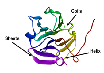
Figure 1. 3D Structure of Hemopexin Like Domain of MMP-2 (PDB ID: 1RTG) Illustrating Coils, Sheets and Helix Region
3.2.Analysis of Physiochemical PropertiesTo analyze physiochemical properties of MMP-2, the ExPASy ProtParam tool was used. This analysis provided key insights into the stability, solubility and characteristics of protein. It showed that hemopexin like domain of MMP-2 protein comprised of 210 amino acids. The instability index was 19.17, indicating that MMP-2 protein is stable as the score less than 40 indicates protein stability. The MMP-2 protein appeared to be hydrophilic based on a negative ground average hydrophobicity gravy coefficient. The protein's pH was represented by the Pi value of 6.81; a value less than 7 suggested that the protein was moderately acidic, which may affect how the protein interacts with the body. The half-life of 20 h suggested that the MMP-2 had moderate stability in a cellular environment as shown in Table 2.
Table 2. The Physiochemical Properties of MMP-2 Protein Obtained Through ExPASy ProtParam
|
S.no |
Instability index |
Gravy Value |
Pi value |
Extinction coefficient |
Half life |
|---|---|---|---|---|---|
|
1 |
19.17 |
-0.305 |
6.81 |
51910 |
20 hours |
By utilizing PSIPRED, the secondary structure of the MMP-2 protein was predicted. Alpha helices and beta sheets were distributed throughout the MMP-2 protein, as seen in Figure 2. Pink region represented 14 residues of alpha helix, 75 residues of yellow region indicated beta sheets, and the rest of the grey part depicted the loop and coil region. Overall results indicated the uniform distribution of alpha and beta helix making it a well-defined functional protein.
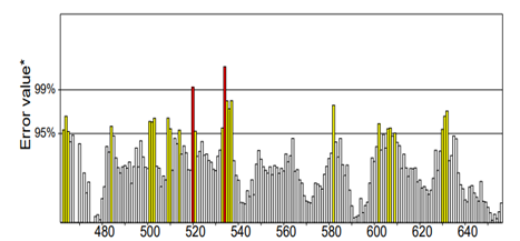
Figure 2. Secondary Structure of MMP-2 Obtained Through PSIPRED
3.4. Analysis of Protein QualityThe ERRAT tool was used to evaluate the overall quality of MMP-2 protein providing an overall quality factor of 86.772 (Figure 3). The analysis indicated that the overall structure was suitable for docking but improvement in certain regions was required, as an ERRAT value of 90 or higher was regarded as an acceptable result.
Figure 3. Results obtained through ERRAT
The Y axis represents the error rate, and the X axis represents the sequence position. Red bars indicate residues where error exceed 99% confidence, yellow bar indicates residues exceeding 95% confidence and white and grey bar represents region with acceptable limits.
3.5.Refinement of the ProteinTo increase protein quality, Galaxy Refine tool was used. The tool worked by enhancing backbone flexibility, optimizing side-chain and minimizing steric clashes. The obtained 97.5 Rama favored value confirmed that the protein had strong geometry leading to more reliable and accurate structure for molecular docking as shown in Table 3.
Table 3. Galaxy Refine Index of MMP-2
|
Model |
GDT-HA |
RMSD |
Mol Probity |
Clash score |
Poor rotamers |
Rama favored |
|---|---|---|---|---|---|---|
|
MODEL 1 |
0.9692 |
0.387 |
1.713 |
12.7 |
0.0 |
97.5 |
After refining the structure of MMP-2 protein, the ERRAT score was checked again, increasing the overall quality factor from 86.772 to 94.086, indicating that the protein was now more stable as the ERRAT score was above 90. The result (Figure 4) showed that the refined structure was of higher quality compared to the initial model shown in Figure 3.
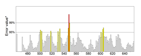
Figure 4. ERRAT results after refining the protein
3.6. Ramachandran PlotTo evaluate the allowed zones of phi and psi dihedral angles in protein structure, a Ramachandran plot was constructed. The analysis revealed that 92.9% of protein residues were in most favorable region, while 5.9% and 1.2% were in generously allowed region, and 0% residue was in disallowed region demonstrating an excellent structural geometry (Figure 5).
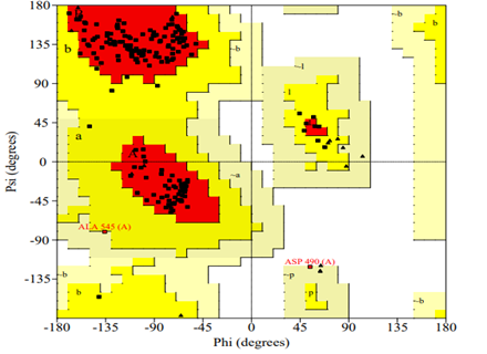
Figure 5. The Ramachandran Plot Findings Reveal That the Most Favorable Zone Contains 92.9% of the Residues
3.7.Binding Affinity of the Cephalosporins to MMP-2Among the eight cephalosporins evaluated, Cefoperazone exhibited the robust molecular interactions with the MMP-2 protein. The most significant binding affinity recorded was −8.1 Kcal/mol for cefoperazone in relation to MMP-2. In contrast, the remaining ligand molecules demonstrated comparatively lower binding affinities, ranging from −5.3 to −6.9 Kcal/mol, as presented in Table 4. The amino acids LYS 489, GLY 634, ARG 567 and LYS 519 contributed in the hydrogen bonding between cefoperazone and MMP-2 as elaborated in Table 5. 3D and 2D representations illustrating the molecular interactions of cefoperazone with MMP-2 are demonstrated in Figure 6 A and B, respectively.
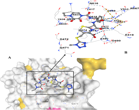
Figure 6. Interaction of Cefoperazone with MMP-2. (A) Cefoperazone (Ligand; Ball and Stick Form) Interacting with MMP-2 (Surface Form). (B) 2D Image Shows Hydrogen Bonds and Hydrophobic Interactions between Cefoperazone and MMP-2. Weak Hydrogen Bond: (Light Blue Lines), Strong Hydrogen Bond (Dark Blue Lines), Cation Pi Interaction (Orange Line) and Hydrophobic Contact (Grey Line)
Table 4. Interaction Energies of Selected Ligands with MMP-2
|
S no. |
Ligand Name |
Interactions Energies (Kcal/mol) |
|---|---|---|
|
1 |
Cefoperazone |
-8.1 |
|
2 |
Cefixime |
-6.9 |
|
3 |
Ceftazidime |
-6.4 |
|
4 |
Ceftizoxime |
-6.4 |
|
5 |
Cefditoren |
-6.3 |
|
6 |
Ceftibuten |
-6.0 |
|
7 |
Cefpodoxime |
-5.9 |
|
8 |
Cefotaxime |
-5.3 |
Table 5. MMP-2 Amino Acids Involved in Different Types of Interactions with Cefoperazone
|
Type of interactions |
Amino acids |
|---|---|
|
Weak Hydrogen bond |
LYS 489, GLY 634 |
|
Strong hydrogen bond |
ARG 567, LYS 519 |
|
Cation pi interaction |
LYS 489, ARG 564 |
|
Hydrophobic contact |
LYS 633 |
In biological macromolecules, flexibility is a crucial property for mediating protein–ligand interactions or for facilitating interactions with substrates. Therefore, iMODS used NMA to calculate molecular motion and geometric flexibility, and then integrated this information with the docked complex coordinates. The output generated by the server illustrated that the deformability of the complex was primarily influenced by the individual distortions of each residue, which were depicted through hinges (highest green peaks) along the chain (Fig. 7A). B-factor values served to quantify the uncertainty associated with each individual atom. The B-factor graph provided a lucid visualization of the relationship between the docked complex, the NMA, and the PDB sector. The B-factor graph specifically represented the stable structure of the docked molecules on the basis of average RMS, as demonstrated in Fig 7B. The server calculated the eigenvalue to be 1.513588e−04, indicating less fluctuation and stabilization of hemopexin domain after binding drug molecule which supported allosteric inhibition mechanism as depicted in Fig. 7D. According to each normal mode, eigenvalue and variance also have an inverse relationship (Fig. 7C). Correlated, uncorrelated, and anti-correlated fluctuations were denoted by red, white, and blue colors, respectively, in the graphic representation of the covariance matrix (Fig. 7E). The docked protein molecule (Cα) atoms were held together by "springs" of different strengths, as shown by an elastic network model. Light grey revealed flexibility, while darker greys demonstrated more rigid springs.
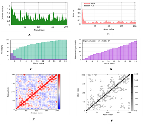
Figure 7. iMODS Normal Mode Analysis (NMA) of Cefoperazone with Targeted MMP-2 Protein Illustrating the Following Graphical Representations: (A) Deformation Plot, (B) B-Factor, (C) Variance Plot, (D) Eigenvalues, (E) Covariance Matrix Plot (Where the Anticorrelated, Uncorrelated, and Correlated States of Atomic Motion are Depicted by Blue, White, and Red Hues, Respectively), and (F) Elastic Network Model, in Which Connections between Atoms are Denoted in Grey
4.DISCUSSION
MMP-2 inhibition for cancer therapy is considered as a great approach as its role has been studied in ovarian cancer. MMP-2 plays a crucial role in both normal physiological processes and the pathology of ovarian cancer. In healthy tissues, it is involved in ECM remodeling, which is essential for tissue repair, wound healing, and angiogenesis. However, its dysregulation is linked to various diseases, including cancer, where it facilitates tumor invasion and metastasis [21]. In ovarian cancer, MMP-2 expression is significantly elevated compared to normal tissues, correlating with advanced disease stages and histological grades [22]. High level of MMP-2 expression has been previously linked with poor prognosis in ovarian cancer patients, making it a viable therapeutic target [23, 24].
Cephalosporins, a class of β-lactam antibiotics, has strong antibacterial effects and has minimum toxicity in humans [25]. Apart from their bactericidal role, their anti-cancer activity has also been studied previously. They can cause DNA damage, mostly by generating reactive oxygen species [26, 27], which can be toxic to cancer cells [28, 29]. This study investigated the potential of cephalosporins to be used as a drug in order to inhibit matrix metalloproteinase-2 in ovarian cancer through in silico drug repurposing and molecular docking.
The initial phase of this study focused on the analysis of the MMP-2 protein. The protein sequence, retrieved from UniProt (P08253), was subjected to physicochemical property analysis with the help of the ExPASy ProtParam tool. Results from this analysis showed that MMP-2 is a stable, hydrophilic protein that had an instability index of 19.17. The visualization of the three dimensional structure of the MMP-2 was retrieved and deposited in the protein data bank (PDB) and visualized in discovery Studio. Secondary structure prediction results obtained through PSIPRED indicated a balanced proportions of alpha helices and beta sheets. This demonstrates that the protein understudy, MMP-2 is a well characterized functional protein. The MMP-2 structure was improved using the Galaxy Refine tool. This step was necessary because geometric discrepancies, missing amino acid residues, steric clashes, and different resolutions in published PDB structures influence docking and simulation analysis [30]. The refining process contributed to a higher score of the ERRAT quality that rose to 94.086 from 86.772 while Ramachandran plot analysis showed that 97.5% of residues were present in favorable regions which confirms the structural integrity of the refined MMP-2 model.
The core of this study focused on evaluating the interactions between MMP-2 and selected cephalosporins. In silico methods like drug repurposing and molecular docking, two promising tools were used to identify potential inhibitors that would selectively restrict the activity of MMP-2 regulatory domain and reducing off target effects linked with zinc ion chelation [31]. Molecular docking studies, which are considered mostly accurate to identify potential inhibitors [32], revealed that cefoperazone exhibited the highest binding affinity, with an interaction energy of -8.1 Kcal/mol. This intrinsic interaction is an indication that cefoperazone can be very useful in the inhibition of MMP-2 activity. Detailed analysis of the cefoperazone-MMP-2 complex indicated specific molecular interactions. All the ligand molecules have shown interactions with MMP-2 with all the molecules showing negative ΔG (binding affinity). In a time-invariant electric field, electrostatic energy is the potential energy of a system; a positive value denotes repulsion, while a negative value denotes surface contact between two molecules. Cefoperazone demonstrated stronger binding affinity with MMP-2 measuring −8.1 ΔG (kcal mol−1).
The negative value of docking complex has confirmed the thermodynamic stability in docked structure. While studying protein-ligand interactions, hydrogen bonding, hydrophobic interactions and electrostatic interactions were considered. The interactions studied between the two molecules showed strong hydrogen bonds with ARG 567 and LYS 519, weak hydrogen bonds with LYS 489 and GLY 634, cation-pi interactions with LYS 489 and ARG 564, and hydrophobic contact with LYS 633. These interactions contribute to the stability and efficacy of the cefoperazone-MMP-2 complex. Thus, after analyzing the ∆G value for cefoperazone and MMP-2 complex by molecular docking, it can be concluded that cefoperazone exhibited highest binding affinity with MMP-2.
The flexibility of the Cefaperazone-MMP-2 complex was investigated using iMODS. The analysis of the internal coordinates based on the structural interactions between proteins and ligands was done by iMODS. B-factor is also attributed to the mobility and deformability of a protein and deformability measures the flexibility of a protein [33]. The generated eigenvalues of the docked proteins are also closely related to the energy required to modify the structure. It describes the restricted motion of the protein-ligand complex. The complex becomes more deformable as the eigenvalue reduces [34]. We discovered that this complex has very low eigenvalue with appreciable degree of deformablity and it indicates that the molecular motion of the docked protein complex was not only flexible but stable as well. Good correlations with slight anticorrelations were depicted by the covariance matrix. Additionally, the elastic network map revealed positive outcomes.
Hemopexin like domain of MMP-2 is an important regulatory domain in recognition of substrates, deactivation of enzyme and contact with metalloproteinase tissue inhibitors (TIMPs) [35, 36]. The observed interactions indicate that Cefoperazone can affect MMP-2 activity via an allosteric inhibition mechanism. Allosteric inhibition of MMP-2 provides a novel promising pathway enhancing selectivity and bypassing the off-target toxicity related with Zn-chelating inhibitors [37, 38]. The hypothesis is also supported by the conformational stabilization during the iMODS analysis. The catalytic-domain structure should be further investigated in the future to uncover the possibility of Cefoperazone binding with Zn2+ center for evaluation of its dual inhibitory potential.
The repurposing of third-generation antibiotics for cancer treatment presents a double-edged sword, as it may cause significant side effects, including the development of antibiotic resistance and off target effects. Modifying existing antibiotics, controlling dose concentration and using combination therapy can improve efficacy while reducing unwanted cellular responses, leading to better survival rates in cancer patients. Another approach is to use nanocarriers like liposomes, dendrimers, and nanoparticles, which allow precise targeting of cancer cells [39].
4.1.ConclusionIn the current investigation, cefoperazone has manifested the most substantial interaction energy with the MMP 2 protein. The current study hypothesizes that an FDA-approved antibiotic cefoperazone can be used in a new therapeutic method, as it directly acts on MMP-2 as a target in ovarian cancer. Repurposing cefoperazone would help develop new therapy solutions in patients with ovarian cancer using its favorable safety profile. The negative ΔG value of the docking complex (-8.1 kcal/mol) further indicates the thermodynamically stable nature of the interaction.
While these in silico results are promising, they represent a preliminary step. Computational predictions require validation through in vitro and in vivo studies to confirm the inhibitory effects of cefoperazone on MMP-2 activity and its impact on ovarian cancer progression. Future research should also investigate the pharmacokinetics and optimal dosing regimens for cefoperazone in an oncological context, as well as potential synergistic effects when combined with existing chemotherapeutic agents.
CONFLICT OF INTEREST
The authors of the manuscript have no financial or non-financial conflict of interest in the subject matter or materials discussed in this manuscript.
DATA AVALIABILITY STATEMENT
The data associated with the study will be provided by the corresponding author if requested.
FUNDING DETAILS
No funding was received for this research.
ACKNOWLEDGMENT
The authors gratefully acknowledge Informatix Biolabs for providing the platform that made this collaboration possible.
REFERENCES
- Bucur C, Balescu I, Petrea S, et al. Ovarian cancer prevention and screening–where do we stand today? J Mind Med Sci. 2024;11(1):99–105. https://doi.org/10.22543/2392-7674.1464
- Bindhu DKR, Murugan L, Mathews SG, et al. Molecular mavericks: unveiling the matrix metalloproteinase gene’s role in ovarian cancer. Egypt J Med Human Genet. 2025;26(1):e65. https://doi.org/10.1186/s43042-025-00693-9
- Murray NPJO. The role of matrix metalloproteinase-2 in the metastatic cascade: a review. Oncol. 2024;26(1):27–40. https://doi.org/10.1515/oncologie-2023-0368
- Assidi M, Jafri MA, Abu-Elmagd M, et al. Prognostic value of matrix metalloproteinase 2 protein expression in ovarian cancer is age-and stage-dependent. Eur J Gynaecol Oncol. 2022;43(1):26–35. https://doi.org/10.31083/j.ejgo4301010
- Jia H, Zhang Q, Liu F, Zhou D. Prognostic value of MMP-2 for patients with ovarian epithelial carcinoma: a systematic review and meta-analysis. Arch Gynecol Obst. 2017;295(3):689–696. https://doi.org/10.1007/s00404-016-4257-9
- Wolosowicz M, Prokopiuk S, Kaminski TW. The complex role of matrix metalloproteinase-2 (MMP-2) in health and disease. Int J Molecul Sci. 2024;25(24):e13691. https://doi.org/10.3390/ijms252413691
- Cavalcante BRR, Freitas RD, da Rocha LOS, et al. In silico approaches for drug repurposing in oncology: a scoping review. Front Pharmacol. 2024;15:e1400029. https://doi.org/10.3389/fphar.2024.1400029
- Sahu R, Rawal RK. Modulation of the c-JNK/p38-MAPK signaling pathway: Investigating the therapeutic potential of natural products in hypertension. Phytomed Plus. 2024;4(2):e100564. https://doi.org/10.1016/j.phyplu.2024.100564
- Jin G, Wong STC. Toward better drug repositioning: prioritizing and integrating existing methods into efficient pipelines. Drug Discover Today. 2014;19(5):637–644. https://doi.org/10.1016/j.drudis.2013.11.005
- Arda AG, Syaifie PH, Ramadhan D, et al. Activity of propolis compounds as potential MMP1 and MMP2 inhibitors by in silico studies in wound healing application. J Pharm Pharm Res. 2024;12(2):264–285. https://doi.org/10.56499/jppres23.1719_12.2.264
- Xia D, Yang X, Liu W, et al. Over-expression of CHAF1A in Epithelial Ovarian Cancer can promote cell proliferation and inhibit cell apoptosis. Biochem Biophy Res Commun. 2017;486(1):191–197. https://doi.org/10.1016/j.bbrc.2017.03.026
- Saeidnia S. New approaches to natural anticancer drugs. Springer Nature; 2015.
- He X, Yao Q, Fan D, et al. Combination of cefotaxime and cisplatin specifically and selectively enhances anticancer efficacy in nasopharyngeal carcinoma. Curr Cancer Drug Targets. 2023;23(7):572–584. https://doi.org/10.2174/1568009623666230227162532
- Ali ZA, Jawad ZN. Anti-Cancer effect of cephalexin loaded on nano chitosan against humane liver cancer. J Pioneer Med Sci. 2024;13:15–17. https://doi.org/10.61091/jpms202413203
- Varghese D, Sunny D, Kurian A, Cherian T, Varghese L. An in silico study on reproposing eravacycline as an MMP inhibitor. J Appl Pharm Sci. 2023;13(1):232–240. https://doi.org/10.7324/japs.2023.130112
- Lee KN, Park KH, Ahn K, Im EM, Oh E, Cho I. Extracellular matrix‐related and serine protease proteins in the amniotic fluid of women with early preterm labor: Association with spontaneous preterm birth, intra‐amniotic inflammation, and microbial invasion of the amniotic cavity. Am J Reproduc Immunol. 2023;90(1):e13736. https://doi.org/10.1111/aji.13736
- Gasteiger E, Hoogland C, Gattiker A, et al. Protein identification and analysis tools on the ExPASy server. In: Walker JM, ed. The Proteomics Protocols Handbook. Springer; 2005:571–607.
- Colovos C, Yeates TO. Verification of protein structures: patterns of nonbonded atomic interactions. Prot Sci. 1993;2(9):1511–1519. https://doi.org/10.1002/pro.5560020916
- Trott O, Olson AJ. AutoDock Vina: improving the speed and accuracy of docking with a new scoring function. J Comput Chem. 2009;31:455–461. https://doi.org/10.1002/jcc.21334
- López-Blanco JR, Aliaga JI, Quintana-Ortí ES, Chacón P. iMODS: internal coordinates normal mode analysis server. Nucleic Acids Res. 2014;42(W1):W271–W276. https://doi.org/10.1093/nar/gku339
- Wolosowicz M, Prokopiuk S, Kaminski TW. The complex role of matrix Metalloproteinase-2 (MMP-2) in Health and Disease. Int J Molecul Sci. 2024;25(24):e13691. https://doi.org/10.3390/ijms252413691
- Liu C. Pathological and prognostic significance of matrix metalloproteinase-2 expression in ovarian cancer: a meta-analysis. Clinical Exper Med. 2016;16(3):375–382. https://doi.org/10.1007/s10238-015-0369-y
- Davidson B, Goldberg I, Gotlieb WH, et al. High levels of MMP-2, MMP-9, MT1-MMP and TIMP-2 mRNA correlate with poor survival in ovarian carcinoma. Clinic Exper Metast. 1999;17(10):799–808. https://doi.org/10.1023/a:1006723011835
- Kenny HA, Lengyel E. MMP-2 functions as an early response protein in ovarian cancer metastasis. Cell Cycle. 2009;8(5):683–688. https://doi.org/10.4161/cc.8.5.7703
- Nath AP, Balasubramanian A. Cephalosporins: an imperative antibiotic over the generations. Int J Res Pharm Sci. 2020;11(1):623–629. https://doi.org/10.26452/IJRPS.V11I1.1866
- Kalghatgi S, Spina CS, Costello JC, et al. Bactericidal antibiotics induce mitochondrial dysfunction and oxidative damage in mammalian cells. Sci Translat Med. 2013;5(192):192ra85–192ra85. https://doi.org/10.1126/scitranslmed.3006055
- Labay E, Mauceri HJ, Efimova EV, et al. Repurposing cephalosporin antibiotics as pro-senescent radiosensitizers. Oncotarget. 2016;7(23):33919–33933. https://doi.org/10.18632/oncotarget.8984
- Zhang Z, Bi C, Fan Y, Wang H, Bao Y. Cefepime, a fourth-generation cephalosporin, in complex with manganese, inhibits proteasome activity and induces the apoptosis of human breast cancer cells. Int J Molecul Med. 2015;36(4):1143–1150. https://doi.org/10.3892/ijmm.2015.2297
- He X, Yao Q, Fan D, et al. Cephalosporin antibiotics specifically and selectively target nasopharyngeal carcinoma through HMOX1-induced ferroptosis. Life Sci. 2021;277:e119457. https://doi.org/10.1016/j.lfs.2021.119457
- Rehman HM, Naz M, Ghous AG, Malik M, Ahmad S, Bashir H. Computational design and evaluation of a novel temporin 1CEa-IL24 fusion protein for anti-tumor potential. Biomed Res Ther. 2025 12(2):7138–7152. https://doi.org/10.15419/bmrat.v12i2.959
- Rotenberg N, Bentolila NY, Shifman JM. Engineered protein inhibitors for precise targeting of matrix metalloproteinases. Trends Biochem Sci. 2025;50(10):919–930. https://doi.org/10.1016/j.tibs.2025.08.002
- Baidya SK, Patel T, Himaja A, et al. Biphenylsulfonamides as effective MMP-2 inhibitors with promising antileukemic efficacy: synthesis, in vitro biological evaluation, molecular docking, and MD simulation analysis. Drug Develop Res. 2024;85(6):e22255. https://doi.org/10.1002/ddr.22255
- Kovacs JA, Chacón P, Abagyan. Predictions of protein flexibility: first‐order measures. Proteins Struct Funct Bioinfo. 2004;56(4):661–668. https://doi.org/10.1002/prot.20151
- Ghosh P, Bhakta S, Bhattacharya M, et al. A novel multi-epitopic peptide vaccine candidate against Helicobacter pylori: in silico identification, design, cloning and validation through molecular dynamics. Int J Pept Res Therap. 2021;27(2):1149–1166. https://doi.org/10.1007/s10989-020-10157-w
- Lauer-Fields JL, Chalmers MJ, Busby SA, Minond D, Griffin PR, Fields GB. Identification of specific hemopexin-like domain residues that facilitate matrix metalloproteinase collagenolytic activity. J Biol Chem. 2009;284(36):24017–24024. https://doi.org/10.1074/jbc.m109.016873
- Wallon UM, Overall CM. The Hemopexin-like Domain (C Domain) of Human Gelatinase A (Matrix Metalloproteinase-2) Requires Ca2+for Fibronectin and Heparin Binding. J Biol Chem. 1997;272(11):7473–7481. https://doi.org/10.1074/jbc.272.11.7473
- Gioia M, Fasciglione GF, Sbardella D, et al. The enzymatic processing of α-dystroglycan by MMP-2 is controlled by two anchoring sites distinct from the active site. PLoS ONE. 2018;13(2):e0192651–e0192651. https://doi.org/10.1371/journal.pone.0192651
- Christophorus Fideluno Adhipandito, Putri D, Eko Aprilianto, Riris Istighfari Jenie, Belal Al-Najjar, Maywan Hariono. Matrix metalloproteinase9 as the protein target in anti-breast cancer drug discovery: an approach by targeting hemopexin domain. Future J Pharm Sci. 2019;5(1)e1. https://doi.org/10.1186/s43094-019-0001-1
- Dudhe PS, Thakare R. Targeted drug delivery in cancer therapy. Int J Adv Res Sci Commun Technol. 2024;4(1):109–124. https://doi.org/10.48175/ijarsct-22617