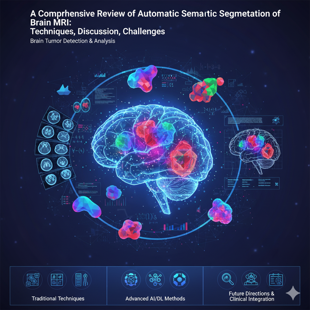
A Comprehensive Review of Automatic Semantic Segmentation of Brain MRI: Techniques, Discussion, Challenges
Abstract
 Abstract Views: 0
Brain tumors, often cancerous, require early detection to improve treatment outcomes and patient survival rates. Automated segmentation of brain tumor images using semantic parameters in medical imaging is complex due to the absence of standardized methods for diverse image dimensions. Challenges include varying image characteristics, disease severity, continuity, content, and non-uniform textures. Clinicians predominantly use labor-intensive manual segmentation. Minimizing user intervention is crucial; current algorithms are primarily semi-automatic, requiring user interaction. Fully automatic methods demand high-resolution MRI images for precise segmentation due to lower noise levels, anatomical consistency, and intensity homogeneity. This review examines traditional and advanced MRI-based segmentation techniques, highlighting challenges, advancements, and proposing future directions for integrating these methods into clinical practice to enhance diagnostic accuracy and treatment efficacy.
Downloads
References
REFERENCES
B. Devkota et al., “Image segmentation for early stage brain tumor detection using mathematical
morphological reconstruction,” Proc. Comput. Sci., vol. 125, pp. 115–123, 2018, doi:
https://doi.org/10.1016/j.procs.2017.12.017.
N. M. Saad, A. Bakar, S. A. R. Sobri Muda, and M. Mokji, “Segmentation of brain lesions in diffusion
weighted MRI using thresholding technique,” presented at the IEEE Int. Conf. Signal Image Process. Appl.,
Kuala Lumpur, Malaysia, Nov. 16–18, 2011, doi: https://doi.org/10.1109/ICSIPA.2011.6144092.
J. Chen et al., “Transunet: Transformers make strong encoders for medical image segmentation,” 2021,
arXiv:2102.04306, doi: https://doi.org/10.48550/arXiv.2102.04306.
A. Kirillov et al., “Segment anything,” presented at 2023 IEEE/CVF International Conference on Computer
Vision (ICCV), Paris, Fracne, 1–6 Oct. 2023, doi: https://doi.org/10.1109/ICCV51070.2023.00371.
N. Mathur, S. Mathur, and D. Mathur, “A novel approach to improve Sobel edge detector,” Proc. Comput. Sci.,
vol. 78, pp. 431–438, 2016, doi: https://doi.org/10.1016/j.procs.2016.07.230.
A. Aslam, E. Khan, and M. M. S. Beg, “Improved edge detection algorithm for brain tumor segmentation,”
Proc. Comput. Sci., vol. 58, pp. 430–437, 2015.
R. D. Nowak, “Wavelet-based Rician noise removal for magnetic resonance imaging,” IEEE Transac. Image
Proc., vol. 8, no. 10, pp. 1408–1419, Oct. 1999, doi: https://doi.org/10.1109/83.791966.
R. Sudharania et al., “Morphological segmentation for brain tumors,” Proc. Comput. Sci., vol. 90, pp. 1–7, 2016.
K. S. A. Viji and J. Jayakumari, “Modified texture based region growing segmentation of MR brain
images,” presented at IEEE Conf. Info. Commun. Technol., Thuckalay, India, April 11–12, 2013, doi:
https://doi.org/10.1109/CICT.2013.6558183.
L. Ling, “Image division method research and realization,” J. Suzhou Coll., vol. 21, pp. 85–88, 2006.
R. Pandav, “Marker-controlled watershed segmentation for brain MRI tumors,” Int. J. Comput. Appl., vol.
, no. 10, pp. 21–27, 2014.
R. Chandra and K. R. H. Rao, “Tumor detection in brain using genetic algorithm,” Proc. Comput. Sci., vol. 79,
pp. 449–457, 2016, doi: https://doi.org/10.1016/j.procs.2016.03.058
J. V. De Oliveira and W. Pedrycz, Advances in Fuzzy Clustering and Its Applications. Hoboken, NJ, USA:
Wiley, 2007.
A. Aina, “Brain tumor segmentation using fuzzy C-means clustering,” Int. J. Comput. Appl., vol. 86, no. 16, pp.
–29, 2014.
K. M. Nimeesha and R. M. Gowda, “Brain tumor segmentation using k-means and fuzzy c-means clustering
algorithm,” IJCSIT Res. Excell., vol. 3, pp. 60–65, Apr. 2013.
B. Tanoori, Z. Azimifar, A. Shakibafar, and S. Katebi, “Brain volumetry: An active contour model-based
segmentation followed by SVM-based classification,” Comput. Biol. Med., vol. 41, pp. 619–632, Aug. 2011,
doi: https://doi.org/10.1016/j.compbiomed.2011.05.013.
S. Bauer, L. P. Nolte, and M. Reyes, “Fully automatic segmentation of brain tumor images using support vector
machine classification in combination with hierarchical conditional random field regularization,” in Medical
Image Computing and Computer-Assisted Intervention, G. Fichtinger, A. Martel, and T. Peters, Eds. Springer,
, pp. 354–361.
H. Al-Shaikhli, R. B. Ahmad, and A. Hussain, “Atlas-based segmentation of brain MRI images,” J. Med. Syst.,
vol. 38, no. 4, p. 33, 2014.
M. Diaz and P. Boulanger, “Atlas-based brain tumor segmentation without deformation model,” Comput Med
Imag Graph, vol. 45, pp. 1–11, 2015.
N. K. Subbanna and T. Arbel, “Probabilistic Gabor and Markov random fields segmentation of brain tumors
in MRI volumes,” in MICCAI-BRATS Workshop, 2012.
N. Subbanna, D. Precup, and T. Arbel, “Iterative multilevel IRF leveraging context and voxel information for
brain tumor segmentation in MRI,” presented at the 2014 IEEE Conf. Computer Vision and Pattern Recognition
(CVPR), Columbus, OH, USA, June 23–28, 2014, doi: https://doi.org/10.1109/CVPR.2014.58.
D. A. Dahab, S. S. A. Ghoniemy, M. Gamal, and A. Selim, “Automated brain tumor detection and identification
using image processing and probabilistic neural network techniques,” Int. J. Image Proc. Visual Commun., vol.
, no. 2, Oct. 2012.
P. A. Mei, C. C. Carneiro, S. J. Fraser, L. L. Min, and F. Reis, “Analysis of neoplastic lesions in magnetic
resonance imaging using self-organizing maps,” J. Neurol. Sci., vol. 359, no. 1-2, pp. 78–83, Dec. 2015, doi:
https://doi.org/10.1016/j.jns.2015.10.032.
A. De and C. Guo, “An adaptive vector quantization approach for image segmentation based on SOM network,”
Neurocomputing, vol. 198, pp. 48–58, Feb. 2015, doi: https://doi.org/10.1016/j.neucom.2014.02.069.
M. Havaei et al., “Brain tumor segmentation with deep neural networks,” Med. Image Anal., vol. 35, pp. 18–31,
Jan. 2017, doi: https://doi.org/10.1016/j.media.2016.05.004.
A. Demirhan and I. Guler, “Combining stationary wavelet transform and self-organizing maps for brain
MR image segmentation,” Eng. Appl. Artif. Intell., vol. 24, pp. 358–356, Mar. 2011, doi:
https://doi.org/10.1016/j.engappai.2010.09.008.
E. A. El-Dahshan et al., “Computer-aided diagnosis of human brain tumor through MRI: A survey and a new
algorithm,” Expert Syst. Appl., vol. 41, no. 11, pp. 5526–5545, Sep. 2014, doi:
https://doi.org/10.1016/j.eswa.2014.01.021.
J. Sachdeva et al., “Sfercb—segmentation, feature extraction, reduction and classification analysis by both
SVM and ANN for brain tumors,” Appl. Soft Comput., vol. 47, pp. 151–167, Oct. 2016, doi:
https://doi.org/10.1016/j.asoc.2016.05.020.
F. Isensee, P. Kickingereder, W. Wick, M. Bendszus, and K. Maier-Hein, “nnu-net: A self-adapting framework
for biomedical image segmentation,” Nat. Meth., vol. 18, pp. 203–211, 2021.
S. Deepak and P. M. Ameer, “Brain tumor classification using deep CNN features via transfer learning,”
Comput. Meth. Prog. Biomed., vol. 111, Art. no. 103345, Aug. 2019, doi:
https://doi.org/10.1016/j.compbiomed.2019.103345.
Z. Ji, Y. Xia, Q. Sun, Q. Chen, and D. Feng, “Adaptive scale fuzzy local Gaussian mixture model for brain MR
image segmentation,” Neurocomputing, vol. 134, pp. 60–69, June 2014, doi:
https://doi.org/10.1016/j.neucom.2012.12.067.
H. Verma, R. K. Agrawal, and A. Sharan, “An improved intuitionistic fuzzy c-means clustering algorithm
incorporating local information for brain image segmentation,” Appl. Soft Comput., vol. 46, pp. 543–557, Sep.
, doi: https://doi.org/10.1016/j.asoc.2015.12.022.
J. Sachdeva et al., “A novel content-based active contour model for brain tumor segmentation,” Magnet.
Resonan. Imag., vol. 30, pp. 694–715, June 2012, doi: https://doi.org/10.1016/j.mri.2012.01.006.
H. H. Sultan, N. M. Salem, and W. Al-Atabany, “Multi-classification of brain tumor images using deep
neural network,” IEEE Access, vol. 7, pp. 69215–69225, 2019, doi:
https://doi.org/10.1109/ACCESS.2019.2919122.
Y. K. Dubey, M. M. Mushrif, and K. Mitra, “Segmentation of brain MR images using rough-set-based
intuitionistic fuzzy clustering,” Biocyber. Biomed. Eng., vol. 36, no. 2, pp. 413–426, 2016, doi:
https://doi.org/10.1016/j.bbe.2016.01.001.
Z. Zhou, M. M. R. Siddiquee, N. Tajbakhsh, and J. Liang, “Unet++: A nested U-Net architecture for medical
image segmentation,” in Deep Learning in Medical Image Analysis and Multimodal Learning for Clinical
Decision Support. Springer Nature, 2018, pp. 3–11.
R. Dubey, M. Hanmandlu, and S. Vasikarla, “Contour-based segmentation with cuckoo search
optimization,” Appl. Soft Comput., vol. 41, pp. 235–247, 2016.
V. Anitha and S. Murugavalli, “Brain tumor classification using two-tier classifier with adaptive segmentation
technique,” IET Computer Vision, vol. 10, no. 1, pp. 9–17, Feb. 2016, doi: https://doi.org/10.1049/iet-
cvi.2014.0193.
N. Tustison et al., “Optimal symmetric multimodal templates and concatenated random forests for supervised
brain tumor segmentation with ANTsR,” Neuroinformatics, vol. 13, no. 2, pp. 209–225, 2015, doi:
https://doi.org/10.1007/s12021-014-9245-2.
L. Huang, S. Ruan, and T. Denœux, “Semi-supervised multiple evidence fusion for brain tumor
segmentation,” Neurocomputing, vol. 535, pp. 40–52, May 2023, doi:
https://doi.org/10.1016/j.neucom.2023.02.047.
K. Srinivas, K. R. Rao, and P. V. G. D. Reddy, “Brain tumor classification using deep neural networks with
feature extraction,” Biomed. Signal Proc. Cont., vol. 72, Art. no. 103285, 2022.
C. Ge, I. Y. H. Gu, A. S. Jakola, and J. Yang, “Deep learning and multi-view fusion for glioma classification
based on multimodal MR imaging,” in Proc. IEEE ISBI, 2018, pp. 411–414.
M. I. Sharif, J. P. Li, M. A. Khan, and M. A. Saleem, “Active deep neural network features selection for
segmentation and recognition of brain tumors using MRI images,” Patt. Recog. Lett., vol. 129, pp. 181–189,
Jan. 2020, doi: https://doi.org/10.1016/j.patrec.2019.11.019.
A. K. Anaraki, M. Ayati, and F. Kazemi, “Magnetic resonance imaging-based brain tumor grades
classification and grading via CNN and genetic algorithm,” Biocyber. Biomed. Eng., vol. 39, no. 1, pp. 63–74,
Jan-Mar. 2019, doi: https://doi.org/10.1016/j.bbe.2018.10.004.
S. Minaee, Y. Boykov, F. Porikli, A. Plaza, N. Kehtarnavaz, and D. Terzopoulos, “Image segmentation using
deep learning: A survey,” IEEE Transac. Patt. Anal. Mach. Intell., vol. 44, no. 7, pp. 3523–3542, July 2022,
doi: https://doi.org/10.1109/TPAMI.2021.3059968.
A. Hatamizadeh, V. Nath, Y. Tang, D. Yang, H. R. Roth, and D. Xu, “UNETR: Transformers for 3D medical
image segmentation,” in Proc. IEEE/CVF Winter Conf. Appl. Comput. Vision, 2022, pp. 574–584.
X. Liu, H. Zhang, Y. Wang, and J. Chen, “Vision transformers for medical image segmentation: A review,”
IEEE Access, vol. 11, pp. 123 456–123 480, 2023.
S. Pereira et al., “Brain tumor segmentation using convolutional neural networks in MRI images,” IEEE
Transac. Med. Imag., vol. 35, no. 5, pp. 1240–1251, 2016.
J. Tang, Z. Li, Q. Chen, and X. Wang, “Optimization of 3d U-Net-based brain tumor segmentation on
consumer-grade edge devices using integer quantization,” ACM Transac. Multimed. Comput. Commun. Appl.,
vol. 20, no. x, pp. 1–20, 2024.
K. Kamnitsas et al., “Efficient multi-scale 3d CNN with fully connected CRF for accurate brain lesion
segmentation,” Med. Image Anal., vol. 36, pp. 61–78, Feb. 2017, doi: https://doi.org/10.1016/j.media.2016.10.004.
R. Wang, P. Liu, S. Zhang, and Y. Li, “Transformer-based brain tumor segmentation in multi-modal mri: A
clinical study,” Human Brain Mapp., vol. 44, no. xx, pp. xxxx–xxxx, 2023.
A. Hatamizadeh, Y. Tang, V. Nath, D. Yang, A. Myronenko, B. Landman, H. R. Roth, and D. Xu, “Swin
UNETR: Swin transformers for 3d medical image segmentation,” in Proc. IEEE/CVF Conf. Comput. Vision
Patt. Recog. Workshps, 2022, pp. 360–371.
A. Myronenko, “3d MRI brain tumor segmentation using autoencoder regularization,” in BrainLes Workshop,
MICCAI, ser. LNCS, vol. 11384, 2018, pp. 311–320.
H. Ye, J. Zhou, and K. Sun, “Deep learning for medical image segmentation: Recent advances and future
trends,” Int. J. Imag. Syst. Technol., vol. 32, no. x, pp. xxx–xxx, 2022.
H. Wang, P. Cao, J. Wang, O. R. Zaiane, and Y. Gao, “TransBTS: Multimodal brain tumor segmentation
using transformer,” IEEE Access, vol. 9, pp. 123 412–123 423, 2021.
P. Afshar, A. Mohammadi, and K. N. Plataniotis, “Brain tumor type classification via capsule networks,” in
Proc. IEEE Int. Conf. Image Proc., 2019, pp. 3129–3133.
M. M. Badzˇa and M. Cˇ. Barjaktarovic´, “Classification of brain tumors from MRI images using a
convolutional neural network,” Appl. Sci., vol. 10, no. 6, 2020, doi: https://doi.org/10.3390/app10061999.
S. U. Khan, M. N. Sharif, M. I. Niass, M. Afzal, and M. Shoaib, “Comparison of multiple deep models on
semantic segmentation for breast tumor detection,” Found. Univ. J. Eng. Appl. Sci., vol. 2, no. 1, pp. 12–23,2021, doi: https://doi.org/10.33897/fujeas.v2i1.424.
X. Gao, Y. Chen, D. Xu, and S. K. Zhou, “Vision transformers in medical image analysis: A comprehensive
survey,” ACM Comput. Surv, vol. 56, pp. 1–39, 2023.
D. Zikic et al., “Decision forests for tissue-specific segmentation of high-grade gliomas in multi-channel MR,” in
Med. Imag Comput. Comput. -Assist. Int., 2012.
S. Rao, S. Vemulapalli, and A. Reddy, “Brain tumor segmentation using random forest and convolutional neural
networks,” in Proc. IEEE Int. Conf. Image Proc., 2015, pp. 335–339.
P. Dvorak and B. Menze, “Structured prediction with CNN features for biomedical image segmentation,” in
Proc. IEEE CVPR Workshops, 2015, pp. 334–342.
G. Urban et al., “Multi-modal brain tumor segmentation using deep convolutional neural networks,” in MICCAI
BraTS Challenge, 2014.
S. U. Khan, F. Wang, J. J. Liou, and Y. Liu, “Segmentation of breast tumors using cutting-edge semantic
segmentation models,” Comput. Meth. Biomech. Biomed. Eng. Imag. Visualiz., vol. 11, no. 2, pp. 242–252, Apr.
, doi: https://doi.org/10.1080/21681163.2022.2064767.
G. Ji, S. Li, and W. Wu, “Brain tumor segmentation using FCM with spatial information,” Comput. Meth.
Program Biomed., vol. 43, no. 10, pp. 1523–1532, 2014.
R. Verma, R. Mehra, and D. Singh, “Brain tumor detection and segmentation using FCM,” Int. J. Adv. Res.
Comput. Sci. Soft. Eng., vol. 5, no. 2, pp. 385–390, 2015.
R. Dubey, M. Hanmandlu, and S. Vasikarla, “Brain tumor detection using intuitionistic rough set-based fuzzy
clustering,” Patt. Recog. Lett., vol. 73, pp. 146–153, 2016.
N. Subbanna, “Markov random field-based segmentation of brain tumors,” Med. Image Anal., vol. 18, no. 2, pp.
–326, 2014.
D. Kwon et al., “Joint segmentation and registration for multifocal gliomas,” IEEE Transac. Med. Imag., vol.
, no. 9, pp. 1873–1885, 2014.
A. Hamamci, N. Kucuk, K. Karaman, and G. Unal, “Tumor-cut: Segmentation of brain tumors on MR images
using a cellular automata method,” Med. Image Anal., vol. 16, no. 4, pp. 766–781, 2012.
K. Thapaliya, J. Y. Pyun, C. S. Park, and G. R. Kwon, “Level set method with automatic selective local
statistics for brain tumor segmentation in MR images,” Comput. Med. Imag. Graph., vol. 37, pp. 522–537,
Dec. 2013, doi: https://doi.org/10.1016/j.compmedimag.2013.05.003.
N. Gordillo, E. Montseny, and P. Sobrevilla, “State of the art survey on MRI brain tumor segmentation,” Mag.
Resonance Imag., vol. 31, no. 8, pp. 1426–1438, Oct. 2013, doi: https://doi.org/10.1016/j.mri.2013.05.002.
A. Isın, C. Direkog˘lu, and M. S¸ ah, “Review of MRI-based brain tumor image segmentation using deep
learning methods,” Proc. Comput. Sci., vol. 102, pp. 317–324, 2016, doi:
https://doi.org/10.1016/j.procs.2016.09.407.
O. Wink, W. J. Niessen, and M. A. Viergever, “Fast delineation and visualization of vessels in 3-d angiographic
images,” IEEE Transac. Med. Imag., vol. 16, pp. 337–346, Apr. 2000, doi: https://doi.org/10.1109/42.848184.
C. Y. Hu, M. D. Grossberg, and G. S. Mageras, “Survey of recent volumetric medical image segmentation
techniques,” in Biomedical Engineering, C. A. B. de Mello, Ed., Intech Open, 2009, pp. 321–346.

Copyright (c) 2025 Sajid Ullah Khan

This work is licensed under a Creative Commons Attribution 4.0 International License.
UMT-AIR follow an open-access publishing policy and full text of all published articles is available free, immediately upon publication of an issue. The journal’s contents are published and distributed under the terms of the Creative Commons Attribution 4.0 International (CC-BY 4.0) license. Thus, the work submitted to the journal implies that it is original, unpublished work of the authors (neither published previously nor accepted/under consideration for publication elsewhere). On acceptance of a manuscript for publication, a corresponding author on the behalf of all co-authors of the manuscript will sign and submit a completed the Copyright and Author Consent Form.


