Kidney Stones: Pathophysiology, Cutting-Edge Treatments, and Emerging Concerns

  • Faiza Saifullah University of Central Punjab, Lahore, Pakistan
  • Muhammad Asad Saeed University of Central Punjab, Lahore, Pakistan
Keywords: calcium oxalate, kidney stones, kidney stone disease, lithotripsy, PCNL, urological complications

Abstract

Abstract Views: 675

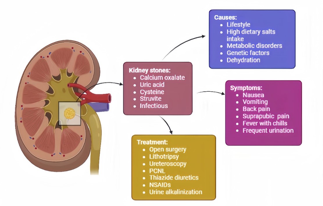
Kidney stone disease (KSD) is a frequently observed clinical condition globally. It poses multiple diagnostic and therapeutic challenges especially in the case of asymptomatic stones when using standard ultrasonic or X-ray procedures. While CT scan options are available for diagnosis, they raise concerns due to excessive radiation exposure. Etiology of KSD is vast and covers disparate types of stones out of which calcium oxalate stones are the most prevalent ones. KSD is influenced by enormous factors ranging from age, gender and obesity to serious metabolic and genetic disorders. Different treatment protocols are available including medications, lithotripsy, ureteroscopy, PCNL and open surgery. Their pros and cons are discussed comprehensively throughout the review. Surgery is only opted for cases when the stone is either extremely large or highly complicated. KSD is a painful condition and most of its treatment options are costly, resulting in a significant financial burden. Despite the availability of advanced treatment protocols, the cases of KSD continue to rise, which is concerning. This suggests that the changing climate and environmental factors which are significantly raising global temperature along with dehydration, may play a role in increasing the prevalence of the disease. The aim of this review is to provide a comprehensive overview of kidney stone disease, providing in-depth insights into stone types and their corresponding management strategies. Furthermore, this review also emphasizes the importance of lifestyle and dietary modifications, identifies constraints observed in the literature survey, and discusses the challenges facing healthcare providers, while also suggesting possible future research directions for emerging studies.

Downloads

Download data is not yet available.

References

Assad A, Raizenne BL, El Yamani MEM, et al. The impact of kidney stone disease on quality of life in high‐risk stone formers. BJU Int. 2024;133(5):570–578. https://doi.org/10.1111/bju.16294

Sorokin I, Mamoulakis C, Miyazawa K, Rodgers A, Talati J, Lotan Y. Epidemiology of stone disease across the world. World J Urol. 2017;35:1301–1320. https://doi.org/10.1007/s00345-017-2008-6

Devi AT, Nagaraj R, Prasad A, Lakkappa DB, Zameer F, Nagalingaswamy NPM. Nephrolithiasis: insights into biomimics, pathogenesis, and pharmacology. Clin Complement Med Pharmacol. 2023;3(2):e100077. https://doi.org/10.1016/j.ccmp.2022.100077

Raheem OA, Khandwala YS, Sur RL, Ghani KR, Denstedt JD. Burden of urolithiasis: trends in prevalence, treatments, and costs. Eur Urol Focus. 2017;3(1):18–26. https://doi.org/10.1016/j.euf.2017.04.001

Sigurjonsdottir VK, Runolfsdottir HL, Indridason OS, Palsson R, Edvardsson VO. Impact of nephrolithiasis on kidney function. BMC Nephrol. 2015;16:1–7. https://doi.org/10.1186/s12882-015-0126-1

Shoag J, Halpern J, Goldfarb DS, Eisner BH. Risk of chronic and end-stage kidney disease in patients with nephrolithiasis. J Urol. 2014;192(5):1440–1445. https://doi.org/10.1016/j.juro.2014.05.117

Khan SR. Nephrocalcinosis in animal models with and without stones. Urol Res. 2010;38:429–438. https://doi.org/10.1007/s00240-010-0303-4

Gambaro G, Croppi E, Bushinsky D, et al. The risk of chronic kidney disease associated with urolithiasis and its urological treatments: a review. J Urol. 2017;198(2):268–273. https://doi.org/10.1016/j.juro.2016.12.135

Uribarri J, Oh MS, Carroll HJ. The first kidney stone. Ann Intern Med. 1989;111(12):1006–1009. https://doi.org/10.7326/0003-4819-111-12-1006

Hyams ES, Matlaga BR. Economic impact of urinary stones. Transl Androl Urol. 2014;3(3):278–283. https://doi.org/10.3978/j.issn.2223-4683.2014.07.02

Faraday M, Hubbard H, Kosiak B, Dmochowski R. Staying at the cutting edge: a review and analysis of evidence reporting and grading; the recommendations of the American Urological Association. BJU Int. 2009;104(3):294–297. https://doi.org/10.1111/j.1464-410X.2009.08729.x

Guyatt GH, Oxman AD, Vist GE, et al. GRADE: an emerging consensus on rating quality of evidence and strength of recommendations. BMJ. 2008;336:e924. https://doi.org/10.1136/bmj.39489.470347.AD

Hughes T, Ho HC, Pietropaolo A, Somani BK. Guideline of guidelines for kidney and bladder stones. Turk J Urol. 2020;46(Suppl 1):S104–S112. https://doi.org/10.5152/tud.2020.20315

Jebir RM, Mustafa YF. Kidney stones: natural remedies and lifestyle modifications to alleviate their burden. Int Urol Nephrol. 2024;56:1025–1033. https://doi.org/10.1007/s11255-023-03764-1

Maline GE, Goldfarb DS. Climate change and kidney stones. Curr Opin Nephrol Hypertens. 2024;33(1):89–96. https://doi.org/10.1097/MNH.0000000000000931

Wei B, Tan W, He S, Yang S, Gu C, Wang S. Association between drinking status and risk of kidney stones among United States adults: NHANES 2007–2018. BMC Public Health. 2024;24:e820. https://doi.org/10.1186/s12889-024-18307-1

Windisch O, Benamran D, Vaessen C. Surgical anatomy of kidneys and adrenals. In: John H, Wiklund P, eds. Robotic Urology. Springer, Cham; 2024:191–101. https://doi.org/10.1007/978-3-031-49428-4_5

Clark‐Price SC, Fischer JR. Physiology, pathophysiology, and anesthetic management of patients with renal disease. In: Lamont L, Grimm K, Robertson S, Love L, Schroeder C, eds. Veterinary Anesthesia and Analgesia: The Sixth Edition of Lumb and Jones. Wiley; 2024:922–943. https://doi.org/10.1002/9781119830306.ch43

Herring LC. Observations on the analysis of ten thousand urinary calculi. J Urol. 1962;88(4):545–562. https://doi.org/10.1016/S0022-5347(17)64842-0

Daudon M, Hennequin C, Lacour B, et al. Sex- and age-related composition of 10,617 calculi analyzed by infrared spectroscopy. Urol Res. 1995;23:319–326. https://doi.org/10.1007/BF00300021

Mandel NS, Mandel GS. Urinary tract stone disease in the United States veteran population. I. Geographical frequency of occurrence. J Urol. 1989;142(6):1513–1515. https://doi.org/10.1016/S0022-5347(17)39144-9

Mandel NS, Mandel GS. Urinary tract stone disease in the United States veteran population. II. Geographical analysis of variations in composition. J Urol. 1989;142(6):1516–1521. https://doi.org/10.1016/S0022-5347(17)39145-0

Pak CY, Poindexter JR, Adams-Huet B, Pearle MS. Predictive value of kidney stone composition in the detection of metabolic abnormalities. Am J Med. 2003;115(1):26–32. https://doi.org/10.1016/S0002-9343(03)00201-8

Pak CY, Kaplan R, Bone H, Townsend J, Waters O. A simple test for the diagnosis of absorptive, resorptive, and renal hypercalciurias. N Engl J Med. 1975;292(10):497–500. https://doi.org/10.1056/NEJM197503062921002

Pacifici R, Rothstein M, Rifas L, et al. Increased monocyte interleukin-1 activity and decreased vertebral bone density in patients with fasting idiopathic hypercalciuria. J Clin Endocrinol Metab. 1990;71(1):138–145. https://doi.org/10.1210/jcem-71-1-138

Pietschmann F, Breslau NA, Pak CY. Reduced vertebral bone density in hypercalciuric nephrolithiasis. J Bone Miner Res. 1992;7(12):1383–1388. https://doi.org/10.1002/jbmr.5650071205

Moe O, Abate N, Sakhaee K. Normouricosuric uric acid urolithiasis: a systemic disease with defective renal acidification. Endocrinol Metab Clin North Am. 2002;31:895–914. https://doi.org/10.1016/S0889-8529(02)00032-4

Malluche HH, Tschoepe W, Ritz E, Meyer-Sabellek W, Massary S. Abnormal bone histology in idiopathic hypercalciuria. J Clin Endocrinol Metab. 1980;50(4):654–658. https://doi.org/10.1210/jcem-50-4-654

Hamm LL, Hering-Smith KS. Pathophysiology of hypocitraturic nephrolithiasis. Endocrinol Metab Clin. 2002;31(4):885–893. https://doi.org/10.1016/S0889-8529(02)00031-2

Pak C, Adams-Huet B, Poindexter J, Pearle M, Peterson R, Moe O. Stone disease rapid communication: Relative effect of urinary calcium and oxalate on saturation of calcium oxalate. Kidney Int. 2004;66:2032–2037.

Cheraghian B, Meysam A, Hashemi SJ, et al. Kidney stones and dietary intake in adults: a population-based study in southwest Iran. BMC Public Health. 2024;24:e955. https://doi.org/10.1186/s12889-024-18393-1

Edvardsson VO, Goldfarb DS, Lieske JC, et al. Hereditary causes of kidney stones and chronic kidney disease. Pediatr Nephrol. 2013;28:1923–1942. https://doi.org/10.1007/s00467-012-2329-z

Beara-Lasic L, Edvardsson VO, Palsson R, Lieske JC, Goldfarb DS, Milliner DS. Genetic causes of kidney stones and kidney failure. Clin Rev Bone Miner Metab. 2012;10:2–18. https://doi.org/10.1007/s12018-011-9113-7

Wu M, Liu M, Zhang Y, et al. Serum HDL partially mediates the association between exposure to volatile organic compounds and kidney stones: A nationally representative cross-sectional study from NHANES. Sci Total Environ. 2024;907:e167915. https://doi.org/10.1016/j.scitotenv.2023.167915

Tamborino F, Cicchetti R, Mascitti M, et al. Pathophysiology and main molecular mechanisms of urinary stone formation and recurrence. Int J Mol Sci. 2024;25(5):e3075. https://doi.org/10.3390/ijms25053075

Dong C, Zhou J, Su X, et al. Understanding formation processes of calcareous nephrolithiasis in renal interstitium and tubule lumen. J Cell Mol Med. 2024;28(7):e18235. https://doi.org/10.1111/jcmm.18235

Coe FL, Parks JH, Asplin JR. The pathogenesis and treatment of kidney stones. N Engl J Med. 1992;327(16):1141–1152. https://doi.org/10.1056/NEJM199210153271607

Devuyst O, Thakker RV. Dent's disease. Orphanet J Rare Dis. 2010;5:e28. https://doi.org/10.1186/1750-1172-5-28

Shrimpton AE, Hoopes RR Jr, Knohl SJ, et al. OCRL1 mutations in Dent 2 patients suggest a mechanism for phenotypic variability. Nephron Physiol. 2009;112(2):27–36. https://doi.org/10.1159/000213506

Tosetto E, Graziotto R, Artifoni L, et al. Dent’s disease and prevalence of renal stones in dialysis patients in Northeastern Italy. J Hum Genet. 2006;51:25–30. https://doi.org/10.1007/s10038-005-0317-x

Diéguez L, Pilco M, Butori S, et al. Dent’s disease: a cause of monogenic kidney stones and nephrocalcinosis. J Pers Med. 2024;14(6):e623. https://doi.org/10.3390/jpm14060623

Wang X, Wang Q. Current dietary and medical prevention of renal calcium oxalate stones. Int J Gen Med. 2024;17:1635–1649. https://doi.org/10.2147/IJGM.S459155

Michael M, Harvey E, Milliner DS, et al. Diagnosis and management of primary hyperoxalurias: best practices. Pediatr Nephrol. 2024;39:3143–3155. https://doi.org/10.1007/s00467-024-06328-2

Cochat P, Rumsby G. Primary hyperoxaluria. N Engl J Med. 2013;369(7):649–658. https://doi.org/10.1056/NEJMra1301564

Bacchetta J, Wood KD. Primary hyperoxaluria type 1: time for prime time? Clin Kidney J. 2022;15:i1–i3. https://doi.org/10.1093/ckj/sfab233

Demoulin N, Aydin S, Gillion V, Morelle J, Jadoul M. Pathophysiology and management of hyperoxaluria and oxalate nephropathy: a review. Am J Kidney Dis. 2022;79(5):717–727. https://doi.org/10.1053/j.ajkd.2021.07.018

Hoppe B. An update on primary hyperoxaluria. Nat Rev Nephrol. 2012;8:467–475. https://doi.org/10.1038/nrneph.2012.113

Abou-Elela A. Epidemiology, pathophysiology, and management of uric acid urolithiasis: a narrative review. J Adv Res. 2017;8(5):513–527. https://doi.org/10.1016/j.jare.2017.04.005

Isaka Y, Takabatake Y, Takahashi A, Saitoh T, Yoshimori T. Hyperuricemia-induced inflammasome and kidney diseases. Nephrol Dial Transplant. 2016;31(6):890–896. https://doi.org/10.1093/ndt/gfv024

Cheungpasitporn W, Thongprayoon C, Mao MA, et al. Incidence of kidney stones in kidney transplant recipients: a systematic review and meta-analysis. World J Transplant. 2016;6(4):790–797. https://doi.org/10.5500/wjt.v6.i4.790

Levy FL, Adams-Huet B, Pak CY. Ambulatory evaluation of nephrolithiasis: an update of a 1980 protocol. Am J Med. 1995;98(1):50–59. https://doi.org/10.1016/S0002-9343(99)80080-1

Bolen E, Stern K, Humphreys M, et al. Urine metabolic risk factors and outcomes of patients with kidney transplant nephrolithiasis. Clin Kidney J. 2022;15(3):500–506. https://doi.org/10.1093/ckj/sfab208

Bunnapradist S, Neri L, Wong W, et al. Incidence and risk factors for diarrhea following kidney transplantation and association with graft loss and mortality. Am J Kidney Dis. 2008;51(3):478–486. https://doi.org/10.1053/j.ajkd.2007.11.013

Rudman D, Dedonis JL, Fountain MT, et al. Hypocitraturia in patients with gastrointestinal malabsorption. N Engl J Med. 1980;303(12):657–661. https://doi.org/10.1056/NEJM198009183031201

Kanbay M, Copur S, Bakir CN, Hatipoglu A, Sinha S, Haarhaus M. Management of de novo nephrolithiasis after kidney transplantation: A comprehensive review from the European Renal Association CKD-MBD working group. Clin Kidney J. 2024;17(2):sfae023. https://doi.org/10.1093/ckj/sfae023

Srivastav S, Guleria K, Sharma S. Predictive machine learning approaches for chronic kidney disease. IEEE. 2023:1–5. https://doi.org/10.1109/INCET57972.2023.10170425

Wu Y, Mo Q, Xie Y, et al. A retrospective study using machine learning to develop predictive model to identify urinary infection stones in vivo. Urolithiasis. 2023;51:e84. https://doi.org/10.1007/s00240-023-01457-z

Doyle P, Gong W, Hsi R, Kavoussi N. PD34-06 machine learning prediction of symptomatic kidney stone recurrence using 24-hour urine data and electronic health record-derived features. J Urol. 2023;209(Suppl 4):e922. https://doi.org/10.1097/JU.0000000000003327.06

Worster A, Preyra I, Weaver B, Haines T. The accuracy of noncontrast helical computed tomography versus intravenous pyelography in the diagnosis of suspected acute urolithiasis: a meta-analysis. Ann Emerg Med. 2002;40(3):280–286. https://doi.org/10.1067/mem.2002.126170

Akram M, Jahrreiss V, Skolarikos A, et al. Urological guidelines for kidney stones: Overview and comprehensive update. J Clin Med. 2024;13(4):e1114. https://doi.org/10.3390/jcm13041114

Frassetto L, Kohlstadt I. Treatment and prevention of kidney stones: an update. Am Fam Physician. 2011;84(11):1234–1242.

Türk C, Petřík A, Sarica K, et al. EAU guidelines on diagnosis and conservative management of urolithiasis. Eur Urol. 2016;69(3):468–474. https://doi.org/10.1016/j.eururo.2015.07.040

Wright P, English P, Hungin A, Marsden S. Managing acute renal colic across the primary-secondary care interface: a pathway of care based on evidence and consensus. BMJ. 2002;325:e1408. https://doi.org/10.1136/bmj.325.7377.1408

Pearle MS, Goldfarb DS, Assimos DG, et al. Medical management of kidney stones: AUA guideline. J Urol. 2014;192(2):316–324. https://doi.org/10.1016/j.juro.2014.05.006

Doyle P, Gong W, Hsi R, Kavoussi N. Machine learning models to predict kidney stone recurrence using 24-hour urine testing and electronic health record-derived features. Res Sq. 2023:rs.3.rs-3107998. https://doi.org/10.21203/rs.3.rs-3107998/v1

Srikanth V. Chronic kidney disease prediction using machine learning algorithms. Int J Techno Eng. 2023;15(1):106–122.

Kaur C, Kumar MS, Anjum A, Binda M, Mallu MR, Al Ansari MS. Chronic kidney disease prediction using machine learning. J Adv Inf Technol. 2023;14(2):384–391. https://doi.org/10.12720/jait.14.2.384-391

Gulhane M, Kumar S, Choudhary S, et al. Integrative approach for efficient detection of kidney stones based on improved deep neural network architecture. SLAS Technol. 2024;29(4):e100159. https://doi.org/10.1016/j.slast.2024.100159

Shee K, Chan C, Liu A, et al. PD34-05 A novel machine-learning algorithm to predict stone recurrence with 24-hour urine data. J Urol. 2023;209(Suppl 4):e921. https://doi.org/10.1097/JU.0000000000003327.05

Anil D, Naimudden S, Reddy AS, Lavanya A. Prediction of chronic kidney disease using various machine learning algorithms. IEEE. 2023:156–161. https://doi.org/10.1109/ICIDCA56705.2023.10100255

Zeng H, Liu Z, He Y, et al. Multivitamins co-intake can reduce the prevalence of kidney stones: a large-scale cross-sectional study. Int Urol Nephrol. 2024;56:2991–3001. https://doi.org/10.1007/s11255-024-04021-9

Hollingsworth JM, Canales BK, Rogers MA, et al. Alpha blockers for treatment of ureteric stones: Systematic review and meta-analysis. BMJ. 2016;355:i6112. https://doi.org/10.1136/bmj.i6112

Bargagli M, Anderegg MA, Fuster DG. Effects of thiazides and new findings on kidney stones and dysglycemic side effects. Acta Physiol. 2024;240(7):e14155. https://doi.org/10.1111/apha.14155

Emiliani E, Subiela JD, Regis F, Angerri O, Palou J. Over 30-year experience on the management of graft stones after renal transplantation. Eur Urol Focus. 2018;4(2):169–174. https://doi.org/10.1016/j.euf.2018.06.007

Normand M, Haymann J-P, Daudon M. Medical treatment of uric acid kidney stones. Can Urol Assoc J. 2024;18(11):E339–345. https://doi.org/10.5489/cuaj.8774

Lam JS, Greene TD, Gupta M. Treatment of proximal ureteral calculi: Holmium:YAG laser ureterolithotripsy versus extracorporeal shock wave lithotripsy. J Urol. 2002;167(5):1972–1976. https://doi.org/10.1016/S0022-5347(05)65065-3

Sharma NKH. A comprehensive review on kidney stones, its treatment and recent advancement. Int J Med Sci Dental Res. 2024;7(1):16–23.

Lingeman JE, Coury TA, Newman DM, et al. Comparison of results and morbidity of percutaneous nephrostolithotomy and extracorporeal shock wave lithotripsy. J Urol. 1987;138(3):485–490. https://doi.org/10.1016/S0022-5347(17)43236-8

Pearle MS, Lingeman JE, Leveillee R, et al. Prospective, randomized trial comparing shock wave lithotripsy and ureteroscopy for lower pole caliceal calculi 1 cm or less. J Urol. 2005;173(6):2005–2009. https://doi.org/10.1097/01.ju.0000158458.51706.56

Torricelli FCM, Danilovic A, Vicentini FC, Marchini GS, Srougi M, Mazzucchi E. Extracorporeal shock wave lithotripsy in the treatment of renal and ureteral stones. Rev Assoc Med Bras. 2015;61(1):65–71. https://doi.org/10.1590/1806-9282.61.01.065

Dasgupta R, Cameron S, Aucott L, et al. Shockwave lithotripsy versus ureteroscopic treatment as therapeutic interventions for stones of the ureter (TISU): a multicentre randomised controlled non-inferiority trial. Eur Urol. 2021;80(1):46–54. https://doi.org/10.1016/j.eururo.2021.02.044

Schlomer BJ. Urologic treatment of nephrolithiasis. Curr Opin Pediatr. 2020;32(2):288–294. https://doi.org/10.1097/MOP.0000000000000849

Swearingen R, Roberts WW, Wolf JS. Ureteroscopy for nephrolithiasis in transplanted kidneys. Can J Urol. 2015;22(2):7727–7731.

Jones P, Elmussareh M, Aboumarzouk OM, Mucksavage P, Somani BK. Role of minimally invasive (micro and ultra-mini) PCNL for adult urinary stone disease in the modern era: evidence from a systematic review. Curr Urol Rep. 2018;19:e27. https://doi.org/10.1007/s11934-018-0764-5

Whitehurst L, Jones P, Somani BK. Mortality from kidney stone disease (KSD) as reported in the literature over the last two decades: a systematic review. World J Urol. 2019;37:759–776. https://doi.org/10.1007/s00345-018-2424-2

Liu M, Chen J, Gao M, et al. Preoperative midstream urine cultures vs renal pelvic urine culture or stone culture in predicting systemic inflammatory response syndrome and urosepsis after percutaneous nephrolithotomy: a systematic review and meta-analysis. J Endourol. 2021;35(10):1467–1478. https://doi.org/10.1089/end.2020.1140

Sharbaugh A, Morgan Nikonow T, Kunkel G, Semins MJ. Contemporary best practice in the management of staghorn calculi. Ther Adv Urol. 2019;11:1–9. https://doi.org/10.1177/1756287219847099

Le Duc A. Les complications immédiates de la chirurgie percutanée du rein [Immediate complications of percutaneous surgery of the kidney]. Prog Urol. 1991;1(1):31–35.

Benchekroun A, Iken A, Karmouni T, et al. La néphrolithotomie percutanée. À propos de 211 cas. Ann Urol. 2001;35(6):315–318. https://doi.org/10.1016/S0003-4401(01)00051-1

Mbodji MM, Jalloh M, Ndoye M, et al. Treatment of kidney stones by percutaneous nephrolithotomy: evaluation of the results of the first series of our experience in the Urology Department of the Idrissa Pouye General Hospital in Dakar. Open J Urol. 2024;14(2):104–114. https://doi.org/10.4236/oju.2024.142011

Wyatt J, Kolettis PN, Burns JR. Treatment outcomes for percutaneous nephrolithotomy in renal allografts. J Endourol. 2009;23(11):1821–1824. https://doi.org/10.1089/end.2008.0115

Krambeck AE, LeRoy AJ, Patterson DE, Gettman MT. Percutaneous nephrolithotomy success in the transplant kidney. J Urol. 2008;180(6):2545–2549. https://doi.org/10.1016/j.juro.2008.08.032

Reddy P, Hulbert J, Lange P, et al. Percutaneous removal of renal and ureteral calculi: experience with 400 cases. J Urol. 1985;134(4):662–665. https://doi.org/10.1016/S0022-5347(17)47372-1

Singh AG, Jairath A, Balaji SS, et al. Changing trends in the endourological management of urolithiasis in anomalous kidneys. BJU Int. 2019;123(2):318–327. https://doi.org/10.1111/bju.14575

Spinos T, Tatanis V, Seitz C, Liatsikos E, Kallidonis P. Percutaneous nephrolithotomy (PCNL) versus other treatments for stone management in horseshoe kidneys: a systematic review. Arab J Urol. 2024;22(4):243–252. https://doi.org/10.1080/20905998.2024.2312699

Boissier R, Rodriguez-Faba O, Zakri RH, et al. Evaluation of the effectiveness of interventions on nephrolithiasis in transplanted kidney. Eur Urol Focus. 2023;9(3):491–499. https://doi.org/10.1016/j.euf.2022.11.019

Wong KA, Olsburgh J. Management of stones in renal transplant. Curr Opin Urol. 2013;23(2):175–179. https://doi.org/10.1097/MOU.0b013e32835d3097

Published
2024-11-15
How to Cite
Saifullah, F., & Saeed, M. A. (2024). Kidney Stones: Pathophysiology, Cutting-Edge Treatments, and Emerging Concerns. Currents in Pharmaceutical Research, 2(2), 89-118. https://doi.org/10.32350/cpr.22.05
Section
Review Article