Convergence of Dermoscopic and Histopathological Findings in Diagnosing Cutaneous Lichen Planus (CLP)
DOI:
https://doi.org/10.32350/BSR.43.03Keywords:
Cutaneous lichen planus (CLP), dermoscopy, diagnosis, histopathology, Wickham striae (WS)Abstract
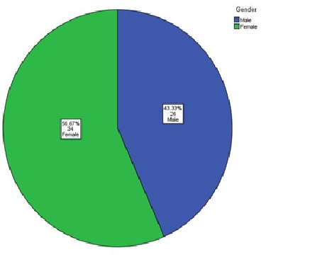
Lichen planus (LP) is an immune mediated disorder that is usually diagnosed clinically. Dermoscopy is a non-invasive diagnostic technique. It can act as an alternative technique to skin biopsy that remains the gold standard for the diagnosis of the disease. The current study aimed to evaluate the degree of convergence between dermoscopy and histopathology in diagnosing LP. It is a cross-sectional study conducted at Jinnah Hospital, Lahore for six (6) months. Sixty (60) patients who met the inclusion criteria were recorded. After taking their informed consent and detailed history, clinical examination and relevant investigations were carried out and recorded in a pre-structured proforma. All patients were subjected to dermoscopic examination. A total of 4 mm punch biopsy were taken from the same site for histopathological examination and sent to JHL Histopathology lab. The results of dermoscopic and histopathological examinations were recorded. Effect of modifiers such as age, gender, and duration of disease were addressed through the stratification of data.Data was analyzed using SPSS 23. The mean age of subjects was 35 years. Of the total 60 patients, 56.6% were female and 43.3% were male. Among the subjects, 94.7% cases diagnosed on histopathology were also diagnosed on dermoscopy, while 5.3% cases were not diagnosed on dermoscopy. Kappa statistics showed a substantial convergence between the two diagnostic modalities: (X2=29.697, p=.000) and (k= .700, p =.000). It was determined that dermoscopy is as effective as hitopathology in the diagnosis of CLP.
Downloads
References
Li C, Tang X, Zheng X, et al. Global prevalence and incidence estimates of oral lichen
planus: A systematic review and meta-analysis. JAMA Dermatol. 2020;156(2):172-181.
https://doi.org/10.1001/jamadermatol.2019.3797
Friedman P, Sabban EC, Marcucci C, Peralta R, Cabo H. Dermoscopic findings in different
clinical variants of lichen planus. Is dermoscopy useful? Dermatol Pract Concept.
;5(4):51-55. https://doi.org/10.5826/dpc.0504a13
Cleach LL, Chosidow O. Lichen Planus. N Engl J Med. 2012;366:723-732.
Gorouhi F, Davari P, Fazel N. Cutaneous and mucosal lichen planus: A comprehensive review
of clinical subtypes, risk factors, diagnosis, and prognosis. Sci World J. 2014;2014:e742826.
https://doi.org/10.1155/2014/742826
Arora SK, Chhabra S, Saikia UN, Dogra S, Minz RW. Lichen planus: A clinical and immuno-
histological analysis. Ind J Dermatol. 2014;59(3):257-261. https://doi.org/10.4103/0019-
131389
Zalaudek I, Lallas A, Moscarella E, Longo C, Soyer HP, Argenziano G. The dermatologist’s
stethoscope—traditional and new application of dermoscopy. Dermatol Pract Concept.
;3(2):e11. http://dx.doi.org/10.5826/dpc.0302a11
Garg P, kaur T, Malhotra SK, Singh A. Study of the dermoscopic findings and their
correlation with histopathological findings in various lichenoid dermatoses. J Clin Exp
Dermatol Res. 2015;6(6):1-8.
Litaiem N, Mansour Y, Jones M, Zeglaoui F. Dermoscopic signs of lichen planus. BMJ Case
Reports 2016. https://doi.org/10.1136/bcr-2015-213923
Gungor S, Topal IO, Goncu EK. Dermoscopic patterns in active and regressive lichen planus
and lichen planus variants: A morphological study. Dermatol Pract Concept. 2015;5(2):45-53.
https://doi.org/10.5826/dpc.0502a06
Lallas A, Kyrgidis A, Tzellos TG, et al. Accuracy of dermoscopic criteria for the diagnosis of
psoriasis, dermatitis, lichen planus and pityriasis roscea. Br J Dermatol. 2012;166(6):1198-
https://doi.org/10.1111/j.1365-2133.2012.10868.x
Jung J, Cho E, Park E, Kim K, Kim K. Atypical dermoscopic findings in patients diagnosed
with lichen planus by histological examination. Dermatologica Sinica. 2017;35(1):20-24.
https://doi.org/10.1016/j.dsi.2016.09.002
Jose S, Kurien G. Diagnostic dermoscopic features and the correlation between dermoscopic
and histopathologic features in lichen planus. Int J Res Dermatol. 2020;6(5):e637.
Downloads
Published
How to Cite
Issue
Section
License
BSR follows an open-access publishing policy and full text of all published articles is available free, immediately upon publication of an issue. The journal’s contents are published and distributed under the terms of the Creative Commons Attribution 4.0 International (CC-BY 4.0) license. Thus, the work submitted to the journal implies that it is original, unpublished work of the authors (neither published previously nor accepted/under consideration for publication elsewhere). On acceptance of a manuscript for publication, a corresponding author on the behalf of all co-authors of the manuscript will sign and submit a completed the Copyright and Author Consent Form.