Veterinary Reproductive Ultrasonography: Advances, Applications, and Future Prospects
DOI:
https://doi.org/10.32350/bsr.73.04Keywords:
diagnosis, imaging, pregnancy, reproduction, veterinary, ultrasonographyAbstract
Background. Ultrasonography is a vital imaging tool in veterinary practice, particularly for reproductive assessment and pregnancy monitoring in animals. Modern ultrasound devices are compact, affordable, and efficient, facilitating their widespread clinical use. Initially applied in livestock for early pregnancy detection in the 1980s, this technique now aids in managing reproductive disorders. Despite its benefits, only a limited number of veterinarians utilize it beyond basic pregnancy diagnosis.
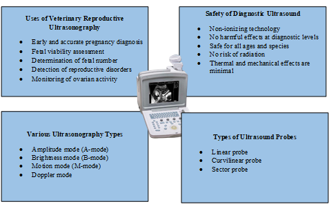
Methods. This review examines the current literature on veterinary reproductive ultrasonography, as well as its various applications, types, modes including A-mode (1D), B-mode (2D), M-mode (motion), and Doppler (blood flow), diagnostic purposes, proper probe selection, patient preparation (hair trimming, gel application), and machine settings. Data from several studies are analyzed to summarize the uses of reproductive ultrasonography in domestic animals.
Results. Ultrasonography enables early pregnancy detection (as early as Day 20-25 in small ruminants and Day 23-30 in cattle), fetal viability assessment, and sex determination (Day 55-60). It accurately monitors ovarian structures (follicles, corpus luteum) and detects reproductive disorders (endometritis, pyometra). Doppler ultrasound evaluates blood flow, while M-mode tracks fetal heart activity. This technique is safe, non-invasive, and enhances the efficiency of reproductive management.
Conclusion. Ultrasonography is a safe, non-invasive, and a highly effective tool for reproductive management in veterinary medicine. It enhances early pregnancy diagnosis, fetal monitoring, and infertility management in livestock and companion animals. Despite its advantages, underutilization persists due to limited expertise among practitioners. Further research is needed to explore its long-term biological effects, though current evidence supports its diagnostic safety and reliability.
Downloads
References
Meomartino L, Greco A, Di Giancamillo M, Brunetti A, Gnudi G. Imaging techniques in veterinary medicine. Part I: radiography and ultrasonography. Eur J Radiol Open. 2021;8:e100382. https://doi.org/10.1016/j.ejro.2021.100382
Mantziaras G, Luvoni GC. Advanced ultrasound techniques in small animal reproduction imaging. Reprod Domest Anim. 2020;55:17–25. https://doi.org/ 10.1111/rda.13587
Lanza GM. Ultrasound imaging: something old or something new? Invest Radiol. 2020;55(9):573–577. https://doi.org10.1097/RLI.0000000000000682
Abdullah R, Asmad K, Khadijah WW, Diyana AN. Ultrasound scanning as a technique in pregnancy diagnosis of Saanen breed at UniSZA dairy goat farm. J Agrobiotechnol. 2018;9(1S):2–11.
Ali Z, Sohail M, Ameen Y, Hamidullah AI, Malik M. Ultrasonography: a tool for management of reproductive disorders in dairy cows. Vet Sci Res Rev. 2023;9(1):18–24. https://doi.org/ 10.17582/journal.vsrr/2023/9.1.18.24
Mali AB, Mehar RM, Kapane SH, Hadimani MR. Scanning the future: veterinary reproductive ultrasonography. Anim Reprod Update. 2022;2(1):82–89. https:// doi.org/10.48165/aru.2022.1201
Ott TL, Tibary A, Waqas M, Geisert R, Giordano J. Pregnancy establishment and diagnosis in livestock. Annu Rev Anim Biosci. 2025;13(1):211–232. http://doi.org/10.1146/annurev-animal-021022-032214
Khand FM, Kachiwal AB, Laghari ZA, et al. Early pregnancy diagnosis and fetometry by real time ultrasonography in teddy goat. J Anim Health Prod. 2021;9(3):342–348. https://doi.org/10. 17582/journal.jahp/2021/9.3.342.348
Bucca S. Use of ultrasonography in fetal development and monitoring. In: Reef VB, Gentile L, Cuervo-Arango J, eds. Atlas of Equine Ultrasonography. Wiley Blackwell; 2022:383–406. https://doi.org/10.1002/9781119514671.ch19
Mali AB, Amle MB, Markandeya NM, Kumawat BL. Comparative efficacy of trans-rectal and trans-abdominal ultrasonography for early diagnosis of pregnancy and embryonic ageing in goats. Indian J Small Rumin. 2019;25(2):171–175. https://doi.org/10.5958/0973-9718.2019.00046.1
Ali A, Derar DR, Abdel-Razek ARK. Ultrasonography for the detection of pregnancy and study of embryonic and fetal development in camels, buffaloes, and sheep: techniques, equations, and limitations. Anim Reprod Sci. 2024;255:e107566. https://doi.org/ 10.1016/j.anireprosci.2024.107566
Özdil A, Yılmaz O. Reproductive doppler ultrasonography practice in bitches. In: Kabu M, Tunç AC, eds. Current Research and Treatment in Animals. 2024;133–147. Livre De Lyon.
Roos J, Aubanel C, Niewiadomska Z, Lannelongue L, Maenhoudt C, Fontbonne A. Triplex Doppler ultrasonography to describe the uterine arteries during diestrus and progesterone profile in pregnant and non-pregnant bitches of different sizes. Theriogenology. 2020;141:153–160. https://doi.org/10.1016/j.theriogenology.2019.08.035
Simon S, Ramanathan A, Ghosh KN. Doppler ultrasonographic assessment of maternal and foetal blood flow in canine pregnancy and its application in the critical management of gestation. Indian J Anim Res. 2022;56(7):811–821. https://doi.org/10.18805/IJAR.B-4286
Saha A, Mathur M. Ultrasound physics & overview. In: Li J, Chow RM-D, Vadivelu N, Kaye AD, eds. Ultrasound Fundamentals: An Evidence-Based Guide for Medical Practitioners. 2021:3–16. https://doi.org/10.1007/978-3-030-46839-2_1
Bagley JE, Richter MP, Lane TJ. The role of transrectal sonography in pregnancy diagnosis in cattle. J Diagn Med Sonogr. 2023;39(1):50–60. https://doi.org/10.1177/87564793221120260
Patey SJ, Corcoran JP. Physics of ultrasound. Anaesth Intensive Care Med. 2021;22(1):58–63. https://doi.org/10.1016/j.mpaic.2020.11.012
Nielsen MB, Søgaard SB, Andersen SB, et al. Highlights of the development in ultrasound during the last 70 years: a historical review. Acta Radiol. 2021;62(11):1499–1514. https://doi.org/10.1177/02841851211050859
Poulose BK. Ultrasonography basics. In: Docimo S, Blatnik JA, Pauli EM, eds. Fundamentals of Hernia Radiology. Cham, Switzerland. Springer International Publishing; 2023:21–27. https://doi.org/10. 1007/978-3-031-21336-6_3
Gjesteby LA, Pare JR, Brattain LJ. Ultrasound for the emergency department and prehospital care. In: Cibis T, McGregor C, eds. Engineering and Medicine in Extreme Environments. Springer International Publishing; 2022:209–234. https://doi.org/10.1007/978-3-030-96921-9_11
Duck FA, Baker AC, Starritt HC, eds. Ultrasound in Medicine. Boca Raton, FL: CRC Press; 2020.
Stewart KA, Navarro SM, Kambala S, et al. Trends in ultrasound use in low and middle income countries: a systematic review. Int J MCH AIDS. 2020;9(1):103–113. https://doi.org/10.21106/ijma.294
Grogan SP, Mount CA. Ultrasound Physics and Instrumentation. StatPearls Publishing; 2023. https://www.ncbi.nlm.nih.gov/books/NBK570593/
Jaśkowski JM, Sobolewski J, Wieczorkiewicz M, Gehrke M, Herudzińska M. Modern techniques of teaching bovine rectal palpation: opportunities, benefits and disadvantages of new educational devices. J Vet Med Educ. 2020;47(1):5–10. https://doi.org /10.3138/jvme.2019-0012
Balaro MFA, Cosentino IO, Ribeiro ACS, Brandão FZ. Ultrasound diagnosis in small ruminants: occurrence and description of genital pathologies. Vet Sci. 2022;9(11):e599. https://doi.org/10.3390/vetsci9110599
Chandolia RK. Applications of transabdominal ultrasonography in bovine reproduction: a review. Buffalo Bull. 2022;41(2):225–240.
Fulton RM. Focused ultrasound of the fetus, female and male reproductive tracts, pregnancy, and dystocia in dogs and cats. Vet Clin North Am Small Anim Pract. 2021;51(6):1249–1265. https://doi.org/10.1016/j.cvsm.2021.07.001
Afzal S, Zahid M, Rehan ZA, et al. Preparation and evaluation of polymer-based ultrasound gel and its application in ultrasonography. Gels. 2022;8(1):e42. https://doi.org/10. 3390/gels8010042
Mattoon JS, Sellon RK, Berry CR. Small Animal Diagnostic Ultrasound. 4th ed. St. Louis, MO: Elsevier; 2020.
Szenci O. Recent possibilities for the diagnosis of early pregnancy and embryonic mortality in dairy cows. Animals. 2021;11(6):e1666. https://doi.org/10.3390/ani11061666
Satheshkumar S. Application of ultrasonography in bovine reproduction. In: Kumaresan A, Srivastava AK, eds. Frontier Technologies in Bovine Reproduction. Singapore: Springer Nature Singapore; 2022:9–45. https://doi.org /10.1007/978-981-19-3072-0_2
Alkan H, Kivrak MB, Satılmış F, Tekindal MA, Dinç DA. Detection of twin pregnancies in ewes by pregnancy-associated glycoprotein assay and transabdominal ultrasonography. Domest Anim Endocrinol. 2020;72:e106399. https://doi.org/10.1016/j.domaniend.2019.106399
Christiansen D. Examination for pregnancy: Rectal palpation. In: Hopper RM, ed. Bovine Reproduction. 2nd ed. Hoboken, NJ: Wiley-Blackwell; 2021:471–478. https:// doi.org/10.1002/9781119602484.ch38
Gardón JC, Acero BÁ, López SR. The use of ultrasonography in female bovine reproduction. In: Gardón JC, Ambrojo KS, eds. Assisted Reproductive Technologies in Animals. Vol 2. Cham, Switzerland: Springer; 2025:3–69. https://doi.org /10.1007/978-3-031-87198-6_1
Adams GP, Singh J. Ovarian follicular and luteal dynamics in cattle. In: Hopper RM, ed. Bovine Reproduction. 2nd ed. Hoboken, NJ: Wiley-Blackwell; 2021:292-323. https:// doi.org/10.1002/9781119602484.ch25
Bajaj NK. Reproduction and kidding in goats. In: Rana T, ed. Trends in Clinical Diseases, Production and Management of Goats. Springer Nature; 2024:151–162. https:// doi.org/10.1016/B978-0-443-23696-9.00019-5
Barrón-Bravo O, Avilés-Ruiz R, Fraga-Escamilla E, Bautista-Martínez Y. Reproductive processes in cows and the ultrasonography use. Abanico Vet. 2023;13:e2023. https://doi.org/ 10.21929/abavet2023.18
Magalhaes HB, Canisso IF, Dell-Aqua JA Jr. The temporal associations of B-mode and power-Doppler ultrasonography, and ovarian steroid changes of the periovulatory follicle and corpus luteum during luteogenesis and luteolysis in jennies. J Equine Vet Sci. 2023;122:e104224. https:// doi.org/10.1016/j.jevs.2023.104224
Yáñez U, Murillo AV, Becerra JJ, Herradón PG, Peña AI, Quintela LA. Comparison between transrectal palpation, B-mode and Doppler ultrasonography to assess luteal function in Holstein cattle. Front Vet Sci. 2023;10:e1162589. https://doi.org /10.3389/fvets.2023.1162589
Sumiyoshi T, Endo N, Tanaka T, Kamomae H. No adverse effect of confirmation of ovulation by rectal palpation and ultrasonography after artificial insemination on formation, development, and function of the corpus luteum and conception rate in cows. J Reprod Dev. 2022;68(6):377–382. https://doi.org/10.1262/jrd.2021-122
Fricke PM. Scanning the future - ultrasonography as a reproductive management tool for dairy cattle. J Dairy Sci. 2002;85(8):1918–1926. https://doi.org/10.3168/jds.S0022-0302(02)74268-9
Gnemmi G, Gardón JC, Maraboli C. Ultrasonography in bovine gynecology. In: Gardón JC, Satué K, eds. Biotechnologies Applied to Animal Reproduction: Current Trends and Practical Applications for Reproductive Management. Boca Raton, FL: Apple Academic Press; 2020:21–40.
Detti L, Francillon L, Christiansen ME, et al. Early pregnancy ultrasound measurements and prediction of first trimester pregnancy loss: a logistic model. Sci Rep. 2020;10(1):e1545. https://doi.org/10.1038/s41598-020-58114-3
Samsar MA, Lakoues HT, Yeddou MN, Aouane ND. Early Pregnancy Diagnosis Methods in Cow [doctoral dissertation]. Algiers, Algeria: Ecole Nationale Supérieure Vétérinaire (ENSV); 2024. http://depot.ensv. dz:8080/jspui/handle/123456789/2909
Ortega-Ferrusola CGA, Da Silva-Álvarez E, Ortiz-Rodríguez JM, et al. Clinical application of Doppler ultrasound in the diagnosis of fertility problems in equines. Rev Bras Reprod Anim. 2021;45(4):459–469. https://doi.org/10.21451/1809-3000.RBRA2021.061
Ortega-Ferrusola C, Gómez-Arrones V, Martín-Cano FE, et al. Advances in the ultrasound diagnosis in equine reproductive medicine: new approaches. Reprod Domest Anim. 2022;57:34–44. https://doi.org /10.1111/rda.14192
Panzani D, Cuervo-Arango J, Fanelli D. Ultrasonographic pregnancy diagnosis in the mare. In: Gardón JC, Ambrojo KS, eds. Assisted Reproductive Technologies in Animals Volume 1: Current Trends for Reproductive Management. Cham, Switzerland: Springer Nature; 2024:29–47. https://doi.org/10.1007/ 978-3-031-73079-5_2
Squires E. Current reproductive technologies impacting equine embryo production. J Equine Vet Sci. 2020;89:e102981. https://doi.org /10.1016/j.jevs.2020.102981
Abdelnaby EA, Alhaider AK, El-Maaty AMA, Ragab RS, Seida AA, El-Badry DA. Ovarian and uterine arteries blood flow velocities waveform, hormones and nitric oxide in relation to ovulation in cows superstimulated with equine chorionic gonadotropin and luteolysis induction 10 and 17 days after ovulation. BMC Vet Res. 2023;19(1):e205. https://doi.org/10. 1186/s12917-023-03692-3
Uçmak ZG, Kurban İ, Uçmak M. The vascularity of preovulatory follicle: the colour-Doppler assessment and its predictive value in the early pregnancy outcome in Arabian mares. Vet Hekim Der Derg. 2020;91(2):104–109. https://doi.org/10.33188/vetheder.672255
Dascanio JJ. Assessment of late-term fetal well-being. In: Dascanio J, McCue P, eds. Equine Reproductive Procedures. Hoboken, NJ: Wiley-Blackwell; 2021:273–276. https:// doi.org/10.1002/9781119556015.ch73
Gao Y, Hannan MA, Murata K, Rajabi-Toustani R, Nambo Y. Ultrasonographic examination of equine fetal growth parameters throughout gestation in pony for equine-assisted therapy. J Vet Med Sci. 2022;84(1):74–81. https://doi.org/ 10.1292/jvms.21-0301
Murase H, Endo Y, Tsuchiya T, et al. Ultrasonographic evaluation of equine fetal growth throughout gestation in normal mares using a convex transducer. J Vet Med Sci. 2014;76(7):947–953. https://doi.org/ 10.1292/jvms.13-0259
Gonzalez SW, Espy BMK, Stefanovski D, Turner RM. Real-time transrectal ultrasonographic measurement of the fetal eye (vitreous body) to predict parturition in bucking horse mares. J Equine Vet Sci. 2025;145:e105345. https://doi.org /10.1016/j.jevs.2025.105345
Lubusky M, Studnickova M, Skrivanek A, Vomackova K, Prochazka M. Ultrasound evaluation of fetal gender at 12-14 weeks. Biomed Pap Med Fac Univ Palacky Olomouc Czech Repub. 2012;156(4):324–329. https://doi.org/10.5507/bp.2012.022
Medan M, Watanabe G, Absy G, et al. Early pregnancy diagnosis by means of ultrasonography as a method of improving reproductive efficiency in goats. J Reprod Dev. 2004;50(4):391–397.
Renaudin C, Wensley F, Morgan J, Cassano J, Spriet M. Transrectal ultrasonographic assessment of the fetal proximal phalanx: a new tool to assess fetal age and bone development in horses. Theriogenology. 2024;226:167–172. https://doi.org /10.1016/j.theriogenology.2024.06.010
Colloton J. Ultrasound evaluation of the female reproductive tract. In: Hopper RM, ed. Bovine Reproduction. 2nd ed. Wiley-Blackwell; 2021:486–508. https://doi.org/10.1002/ 9781119602484.ch40
Medan MS, Abd El-Aty AM. Advances in ultrasonography and its applications in domestic ruminants and other farm animals reproduction. J Adv Res. 2010;1(2):123–128.
Kim D, Son M, Jung D, Heo S, Kim M, Yi J. Economic impacts of ultrasonographic fetal sex determination on Hanwoo cattle profitability and market dynamics. Vet Sci. 2025;12(3):e201. https://doi.org 10.3390/vetsci12030201
Fontes PL, Oosthuizen N. Applied use of Doppler ultrasonography in bovine reproduction. Front Anim Sci. 2022;3:e912854. https://doi.org /10.3389/fanim.2022.912854
Rehman HU, Kanwal M, Zardari AA, et al. Ultrasonographic estimation of fetal gestational age in the second and third trimesters using various biometric parameters. J Popul Ther Clin Pharmacol. 2024;31(5):2121–2126. https://doi.org/10.53555 /jptcp.v31i5.7474
Francis YM, Karunakaran B. Ultrasonographic estimation of the gestational age using the fetal kidney length in the second and third trimesters of pregnancy among South Indian antenatal women: a cross-sectional study. Cureus. 2023;15(6):e40032. https://doi.org/10. 7759/cureus.41172
Published
How to Cite
Issue
Section
License
BSR follows an open-access publishing policy and full text of all published articles is available free, immediately upon publication of an issue. The journal’s contents are published and distributed under the terms of the Creative Commons Attribution 4.0 International (CC-BY 4.0) license. Thus, the work submitted to the journal implies that it is original, unpublished work of the authors (neither published previously nor accepted/under consideration for publication elsewhere). On acceptance of a manuscript for publication, a corresponding author on the behalf of all co-authors of the manuscript will sign and submit a completed the Copyright and Author Consent Form.