
Evaluation of Immune Evasive and Infectivity Potential of Viral Molecular Evolution of SARS-CoV-2 Emerging Variants
DOI:
https://doi.org/10.32350/sir.102.03Keywords:
hACE2, immune evasion, infectivity potential, SARS-CoV-2, spike protein, variants of concern (VOCs)Abstract
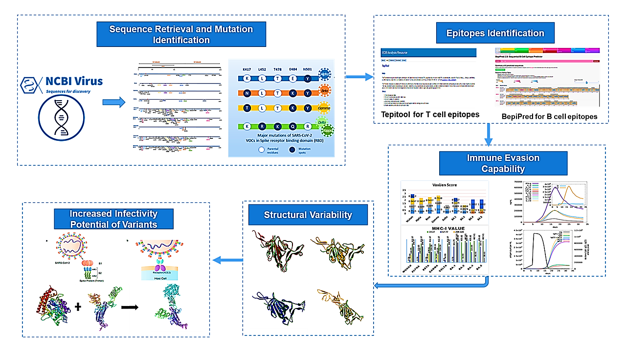
In December 2019, the world was shocked by a new and highly infectious pathogen named SARS-CoV-2 which caused a very high number of cases and deaths, worldwide. Several vaccines were introduced to tackle the virus but complications arose when its different variants were identified. World Health Organization (WHO) has classified them as “Variants of Concern” and “Variants of Interest”. VOCs include Alpha (B.1.17), Beta (B.1.351), Gamma (P.1), Delta (B.1.617), and Omicron (BA.1, BA.2, BA.4 and BA.5). The primary focus of this study was the mutations reported in the spike protein of these VOCs and their role in immune evasion and infectivity potential of the virus. This study employed an in silico approach to provide the detailed structure of the spike protein of all variants. The immune threshold of the VOCs and Wuhan-ref was compared. Further, differences in the interaction of the spike glycoprotein of the variants and Wuhan-ref with the hACE2 receptor were analyzed to show variations in the spike protein of different variants. The findings indicate that as the number of mutations increased in the emerging variants, so did their infectivity potential. Furthermore, when different epitopes were examined for immune evasion, it was found that molecular evolution has made them more immune evasive but less severe than the Wuhan-ref.
Downloads
References
1. Malik YS, Sircar S, Bhat S, et al. Emerging novel coronavirus (2019-nCoV)—current scenario, evolutionary perspective based on genome analysis and recent developments. Vet Q. 2020;40(1):68–76. https://doi.org/10.1080/01652176.2020.1727993
2. Wu F, Zhao S, Yu B, et al. A new coronavirus associated with human respiratory disease in China. Nature. 2020;579(7798):265–269. https://doi.org/10.1038/s41586-020-2008-3
This study reports the identification and characterization of a novel coronavirus associated with human respiratory disease in China. It provides foundational evidence regarding the emergence and genetic characteristics of SARS-CoV-2 and establishes an important basis for subsequent research on viral evolution, infectivity, and pathogenicity.
3. Sanders JM, Monogue ML, Jodlowski TZ, Cutrell JB. Pharmacologic treatments for coronavirus disease 2019 (COVID-19): A review. JAMA. 2020;323(18):1824–1836. https://doi.org/10.1001/jama.2020.6019
4. Paules CI, Marston HD, Fauci AS. Coronavirus infections—more than just the common cold. JAMA. 2020;323(8):707–708. https://doi.org/10. 1001/jama.2020.0757
5. Romano M, Ruggiero A, Squeglia F, Maga G, Berisio R. A structural view of SARS-CoV-2 RNA replication machinery: RNA synthesis, proofreading and final capping. Cells. 2020;9(5):e1267. https://doi.org/10.3390/cells9051267
6. Finkel Y, Mizrahi O, Nachshon A, et al. The coding capacity of SARS-CoV-2. Nature. 2021;589(7840):125–130. https://doi.org/10.1038/ s41586-020-2739-1
7. Jin X, Xu K, Jiang P, et al. Virus strain from a mild COVID-19 patient in Hangzhou represents a new trend in SARS-CoV-2 evolution potentially related to furin cleavage site. Emerg Microbes Infect. 2020;9(1):1474–1488. https://doi.org/10.1080/22221751.2020.1781551
8. Poland GA, Bass J, Goldstein MR. SARS-CoV-2 infections: An ACE in the hole and systems biology studies—a research agenda. Mayo Clin Proc. 2020;95(9):1838–1841. https://doi.org/10.1016/j.mayocp. 2020.06.044
9. Yan R, Zhang Y, Li Y, Xia L, Guo Y, Zhou Q. Structural basis for the recognition of SARS-CoV-2 by full-length human ACE2. Science. 2020;367(6485):1444–1448. https://doi.org/10.1126/science.abb2762
This study investigates the structural basis for the recognition of SARS-CoV-2 by the human ACE2 receptor. Its findings provide important insights into the molecular interaction between the viral spike protein and ACE2, helping explain mechanisms underlying viral entry and infectivity.
10. Haddad D, John SE, Mohammad A, et al. SARS-CoV-2: Possible recombination and emergence of potentially more virulent strains. PLoS One. 2021;16(5):e0251368. https://doi.org/10.1371/journal.pone. 0251368
11. Liu Y, Rocklöv J. The reproductive number of the Delta variant of SARS-CoV-2 is far higher compared to the ancestral SARS-CoV-2 virus. J Travel Med. 2021;28(7):etaab124. https://doi.org/10. 1093/jtm/taab124
12. Planas D, Veyer D, Baidaliuk A, et al. Reduced sensitivity of SARS-CoV-2 variant Delta to antibody neutralization. Nature. 2021;596(7871):276–280. https://doi.org/10.1038/s41586-021-03777-9
13. Karim SSA, Karim QA. Omicron SARS-CoV-2 variant: A new chapter in the COVID-19 pandemic. Lancet. 2021;398(10317):2126–2128. https://doi.org/10.1016/S0140-6736(21)02758-6
14. World Health Organization. Tracking SARS-CoV-2 variants. World Health Organization. Updated July 28, 2026. Accessed September 15, 2026. https://www.who.int/activities/tracking-SARS-CoV-2-variants
15. Rambaut A, Holmes EC, O’Toole Á, et al. A dynamic nomenclature proposal for SARS-CoV-2 lineages to assist genomic epidemiology. Nat Microbiol. 2020;5(11):1403–1407. https://doi.org/10.1038/s41564-020-0770-5
16. Shu Y, McCauley J. GISAID: Global initiative on sharing all influenza data—from vision to reality. Euro Surveill. 2017;22(13):e30494. https://doi.org/10.2807/1560-7917.ES.2017.22.13.30494
17. Ozono S, Zhang Y, Ode H, et al. SARS-CoV-2 D614G spike mutation increases entry efficiency with enhanced ACE2-binding affinity. Nat Commun. 2021;e12:848. https://doi.org/10.1038/s41467-021-21118-2
18. Yurkovetskiy L, Wang X, Pascal KE, et al. Structural and functional analysis of the D614G SARS-CoV-2 spike protein variant. Cell. 2020;183(3):739–751.e8. https://doi.org/10.1016/j.cell.2020.09.032
19. Volz E, Hill V, McCrone JT, et al. Evaluating the effects of SARS-CoV-2 spike mutation D614G on transmissibility and pathogenicity. Cell. 2021;184(1):64–75.e11. https://doi.org/10.1016/j.cell.2020.11.020
20. Mohandas S, Shete A, Kumar A, et al. Comparative pathogenicity of BA.2.12, BA.5.2 and XBB.1 with the Delta variant in Syrian hamsters. Front Microbiol. 2023;14:e1183763. https://doi.org/10.3389/ fmicb.2023.1183763
21. Gruell H, Vanshylla K, Tober-Lau P, et al. Neutralisation sensitivity of the SARS-CoV-2 omicron BA.2.75 sublineage. Lancet Infect Dis. 2022;22(10):1422–1423. https://doi.org/10.1016/S1473-3099(22) 00580-1
22. Baum A, Fulton BO, Wloga E, et al. Antibody cocktail to SARS-CoV-2 spike protein prevents rapid mutational escape seen with individual antibodies. Science. 2020;369(6506):1014–1018. https://doi.org/10. 1126/science.abd0831
23. Chen RE, Zhang X, Case JB, et al. Resistance of SARS-CoV-2 variants to neutralization by monoclonal and serum-derived polyclonal antibodies. Nat Med. 2021;27(4):717–726. https://doi.org/10.1038/ s41591-021-01294-w
24. Liu Z, VanBlargan LA, Bloyet LM, et al. Identification of SARS-CoV-2 spike mutations that attenuate monoclonal and serum antibody neutralization. Cell Host Microbe. 2021;29(3):477–488.e4. https://doi. org/10.1016/j.chom.2021.01.014
25. Liu Y, Liu J, Xia H, et al. Neutralizing activity of BNT162b2-elicited serum. N Engl J Med. 2021;384(15):1466–1468. https://doi.org/10. 1056/NEJMc2102017
26. Brister JR, Ako-Adjei D, Bao Y, Blinkova O. NCBI viral genomes resource. Nucleic Acids Res. 2015;43(D1):D571–D577. https://doi.org/ 10.1093/nar/gku1207
27. Jespersen MC, Peters B, Nielsen M, Marcatili P. BepiPred-2.0: Improving sequence-based B-cell epitope prediction using conformational epitopes. Nucleic Acids Res. 2017;45(W1):W24–W29. https://doi.org/10.1093/nar/gkx346
28. Doytchinova IA, Flower DR. VaxiJen: A server for prediction of protective antigens, tumour antigens and subunit vaccines. BMC Bioinformatics. 2007;8:e4. https://doi.org/10.1186/1471-2105-8-4
29. Paul S, Sidney J, Sette A, Peters B. TepiTool: A pipeline for computational prediction of T cell epitope candidates. Curr Protoc Immunol. 2016;114:18.9.1–18.9.24. https://doi.org/10.1002/cpim.12
30. Rapin N, Lund O, Bernaschi M, Castiglione F. Computational immunology meets bioinformatics: The use of prediction tools for molecular binding in the simulation of the immune system. PLoS One. 2010;5(4):e9862. https://doi.org/10.1371/journal.pone.0009862
31. Gupta R, Jung E, Gooley AA, Williams KL, Brunak S, Hansen J. Scanning the available Dictyostelium discoideum proteome for O-linked GlcNAc glycosylation sites using neural networks. Glycobiology. 1999;9(10):1009–1022. https://doi.org/10.1093/glycob/9.10.1009
32. Gupta R, Brunak S. Prediction of glycosylation across the human proteome and the correlation to protein function. Pac Symp Biocomput. 2002:310–322. https://doi.org/10.1142/9789812799623_0029
33. Blom N, Gammeltoft S, Brunak S. Sequence and structure-based prediction of eukaryotic protein phosphorylation sites. J Mol Biol. 1999;294(5):1351–1362. https://doi.org/10.1006/jmbi.1999.3310
34. Blom N, Sicheritz-Pontén T, Gupta R, Gammeltoft S, Brunak S. Prediction of post-translational glycosylation and phosphorylation of proteins from the amino acid sequence. Proteomics. 2004;4(6):1633–1649. https://doi.org/10.1002/pmic.200300771
35. Schwede T, Kopp J, Guex N, Peitsch MC. SWISS-MODEL: An automated protein homology-modeling server. Nucleic Acids Res. 2003;31(13):3381–3385. https://doi.org/10.1093/nar/gkg520
36. Berman HM, Battistuz T, Bhat TN, et al. The Protein Data Bank. Acta Crystallogr D Biol Crystallogr. 2002;58(Pt 6 No 1):899–907. https://doi.org/10.1107/S0907444902003451
37. Kozakov D, Hall DR, Xia B, et al. The ClusPro web server for protein-protein docking. Nat Protoc. 2017;12(2):255–278. https://doi.org/10. 1038/nprot.2016.169
38. Dassault Systèmes BIOVIA. Pharmacophore and Ligand-Based Design With BIOVIA Discovery Studio. Dassault Systèmes; 2014. Accessed September 15, 2026. https://www.3ds.com/ fileadmin/PRODUCTS-SERVICES/BIOVIA/PDF/Pharmacophore-Ligand-based-Design-with-BIOVIA-Discovery-Studio.pdf
39. Konagurthu AS, Whisstock JC, Stuckey PJ, Lesk AM. MUSTANG: A multiple structural alignment algorithm. Proteins. 2006;64(3):559–574. https://doi.org/10.1002/prot.20921
40. Naz A, Shahid F, Butt TT, Awan FM, Ali A, Malik A. Designing multi-epitope vaccines to combat emerging coronavirus disease 2019 (COVID-19) by employing immuno-informatics approach. Front Immunol. 2020;11:e1663. https://doi.org/10.3389/fimmu.2020.01663
41. Andreano E, Piccini G, Licastro D, et al. SARS-CoV-2 escape in vitro from a highly neutralizing COVID-19 convalescent plasma. bioRxiv. Preprint posted December 28, 2020. https://doi.org/10.1101/ 2020.12.28.424451
42. McCarthy KR, Rennick LJ, Nambulli S, et al. Recurrent deletions in the SARS-CoV-2 spike glycoprotein drive antibody escape. Science. 2021;371(6534):1139–1142. https://doi.org/10.1126/science.abf6950
43. Stokes EK, Zambrano LD, Anderson KN, et al. Coronavirus disease 2019 case surveillance—United States, January 22–May 30, 2020. MMWR Morb Mortal Wkly Rep. 2020;69(24):759–765. https:// doi.org/10.15585/mmwr.mm6924e2
44. Murugesan M, Mathews P, Paul H, Karthik R, Mammen JJ, Rupali P. Protective effect conferred by prior infection and vaccination on COVID-19 in a healthcare worker cohort in South India. PLoS One. 2022;17(5):e0268797. https://doi.org/10.1371/journal.pone.0268797
45. Menni C, Valdes AM, Polidori L, et al. Symptom prevalence, duration, and risk of hospital admission in individuals infected with SARS-CoV-2 during periods of omicron and delta variant dominance: A prospective observational study from the ZOE COVID Study. Lancet. 2022;399(10335):1618–1624. https://doi.org/10.1016/S0140-6736(22) 00327-0
46. Stolp B, Stern M, Ambiel I, et al. SARS-CoV-2 variants of concern display enhanced intrinsic pathogenic properties and expanded organ tropism in mouse models. Cell Rep. 2022;38(7):e110387. https://doi.org/10.1016/j.celrep.2022.110387
47. Liu L, Iketani S, Guo Y, et al. Striking antibody evasion manifested by the Omicron variant of SARS-CoV-2. Nature. 2022;602(7898):676–681. https://doi.org/10.1038/s41586-021-04388-0
This study examines the antibody evasion properties of the Omicron variant of SARS-CoV-2. The findings demonstrate substantial changes in antibody susceptibility associated with Omicron, providing evidence for the role of viral evolution in immune escape.
48. Moriyama M, Lucas C, Monteiro VS, Yale SARS-CoV-2 Genomic Surveillance Initiative, Iwasaki A. SARS-CoV-2 Omicron subvariants evolved to promote further escape from MHC-I recognition. bioRxiv. Preprint originally posted May 4, 2022; updated December 23, 2022. https://doi.org/10.1101/2022.05.04.490614
49. Guo K, Barrett BS, Morrison JH, et al. Interferon resistance of emerging SARS-CoV-2 variants. Proc Natl Acad Sci U S A. 2022;119(32):e2203760119. https://doi.org/10.1073/pnas.2203760119
50. Ledford H. How severe are Omicron infections? Nature. 2021;600(7890):577–578. https://doi.org/10.1038/d41586-021-03794-8
51. Schoenborn JR, Wilson CB. Regulation of interferon-γ during innate and adaptive immune responses. Adv Immunol. 2007;96:41–101. https://doi.org/10.1016/S0065-2776(07)96002-2
52. Schroder K, Hertzog PJ, Ravasi T, Hume DA. Interferon-γ: An overview of signals, mechanisms and functions. J Leukoc Biol. 2004;75(2):163–189. https://doi.org/10.1189/jlb.0603252
53. Liu H, Zhang Q, Wei P, et al. The basis of a more contagious 501Y.V1 variant of SARS-CoV-2. Cell Res. 2021;31(6):720–722. https://doi.org/10.1038/s41422-021-00496-8
54. Raghu D, Hamill P, Banaji A, McLaren A, Hsu YT. Assessment of the binding interactions of SARS-CoV-2 spike glycoprotein variants. J Pharm Anal. 2022;12(1):58–64. https://doi.org/10.1016/j.jpha.2021.09.006
55. Suleman M, Yousafi Q, Ali J, et al. Bioinformatics analysis of the differences in the binding profile of the wild-type and mutants of the SARS-CoV-2 spike protein variants with the ACE2 receptor. Comput Biol Med. 2021;138:e104936. https://doi.org/10.1016/j.compbiomed. 2021.104936
Downloads
Published
How to Cite
Issue
Section
License
Copyright (c) 2026 Nayyer Saddique, Ishaq N. Khan, Jalal Ahmed, Hafsah Muhammad, Muhammad Ibrahim Rashid

This work is licensed under a Creative Commons Attribution 4.0 International License.